

The UMHC Dermatopathology Molecular Diagnostic Laboratory (DPML) offers state of the art molecular diagnostic testing for melanocytic neoplasms and other solid tumors. The tests are intended to aid in the more precise diagnosis of histologically ambiguous neoplasms. This category of atypical lesions cannot be definitively classified as benign or malignant using histopathological criteria alone. Such lesions are usually diagnosed using terms that imply various degrees of uncertainty in regard to their malignant potential and create the possibility for mismanagement including either over or under treatment. Molecular analysis may allow for more precise risk prognostication in these challenging lesions, avoiding unnecessarily aggressive treatment for low-risk lesions, and providing support for appropriate surgical management and staging for high-risk lesions.
Currently, DPML offers 2 tests to aid in the diagnosis of histologically ambiguous melanocytic tumors, a multiprobe FISH assay, and a SNP Chromosomal Microarray assay. Both tests are optimized for formalin-fixed, paraffin-embedded tissue from small biopsies. For both tests, a positive result favors a diagnosis of melanoma while a negative result one of nevus.
The multiprobe FISH assay is designed to detect copy number abnormalities at probed loci via fluorescence in situ hybridization (FISH) in FFPE tissue specimens. Three multiprobe FISH panels are available. Panel 1 include probes 6p25 (RREB1), 6q23 (MYB), 11q13 (CCND1) and CEP6; panel 2 include 9q21 (CDKN2A) and CEP9; panel 3 include 8q24 (MYC). Panel 1 is recommended for all histologically ambiguous melanocytic tumors. The addition of panels 2 and 3 increases assay sensitivity for selected melanocytic neoplasms including spitzoid tumors and nevoid melanomas.
SNP microarray analysis is performed using the ThermoFisher OncoScan (TM) FFPE Assay Kit. The assay utilizes a Molecular Inversion Probe (MIP) technology, which is optimized for highly degraded FFPE samples (probe interrogation site of just 40 base pairs). For copy number analysis the assay has a resolution of 50-100 kb in genomic regions spanning selected 900 cancer genes and of 300 kb outside of these regions.
Besides melanocytic tumors, our lab also offers another 2 tests to aid in the diagnosis and prognosis of other types of tumors FISH for malignancy and SNP Chromosomal Microarray Analysis-Tumor.
FISH for malignancy is designated as single probe FISH for CDKN2A, MYC, or BAP1. The purpose of these tests is to aid in the diagnosis and prognosis of selected neoplasms including atypical vascular lesions (MYC), BAP1-inactivated tumors such as melanocytic tumors or mesothelioma (BAP1) and tumors characterized by homozygous CDKN2A deletion such as mesothelioma.
SNP Chromosomal Microarray Analysis-Tumor is designated to aid in the diagnosis of copy number variations (CNVs) associated tumors. Both tests are optimized for formalin-fixed, paraffin-embedded tissue from small biopsies.
Specimens submitted to the Dermatopathology Molecular Diagnostic Laboratory must be properly labeled and accompanied by a completed DPML Test Requisition. Please refer to the MLabs Test Catalog for available assays and specimen collection requirements.
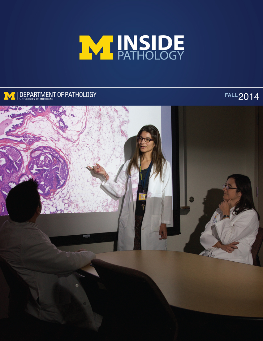 ON THE COVER
Breast team reviewing a patient's slide. (From left to right) Ghassan Allo, Fellow; Laura Walters, Clinical Lecturer; Celina Kleer, Professor. See Article 2014Department Chair |

newsletter
INSIDE PATHOLOGYAbout Our NewsletterInside Pathology is an newsletter published by the Chairman's Office to bring news and updates from inside the department's research and to become familiar with those leading it. It is our hope that those who read it will enjoy hearing about those new and familiar, and perhaps help in furthering our research. CONTENTS
|
 ON THE COVER
Autopsy Technician draws blood while working in the Wayne County morgue. See Article 2016Department Chair |

newsletter
INSIDE PATHOLOGYAbout Our NewsletterInside Pathology is an newsletter published by the Chairman's Office to bring news and updates from inside the department's research and to become familiar with those leading it. It is our hope that those who read it will enjoy hearing about those new and familiar, and perhaps help in furthering our research. CONTENTS
|
 ON THE COVER
Dr. Sriram Venneti, MD, PhD and Postdoctoral Fellow, Chan Chung, PhD investigate pediatric brain cancer. See Article 2017Department Chair |

newsletter
INSIDE PATHOLOGYAbout Our NewsletterInside Pathology is an newsletter published by the Chairman's Office to bring news and updates from inside the department's research and to become familiar with those leading it. It is our hope that those who read it will enjoy hearing about those new and familiar, and perhaps help in furthering our research. CONTENTS
|
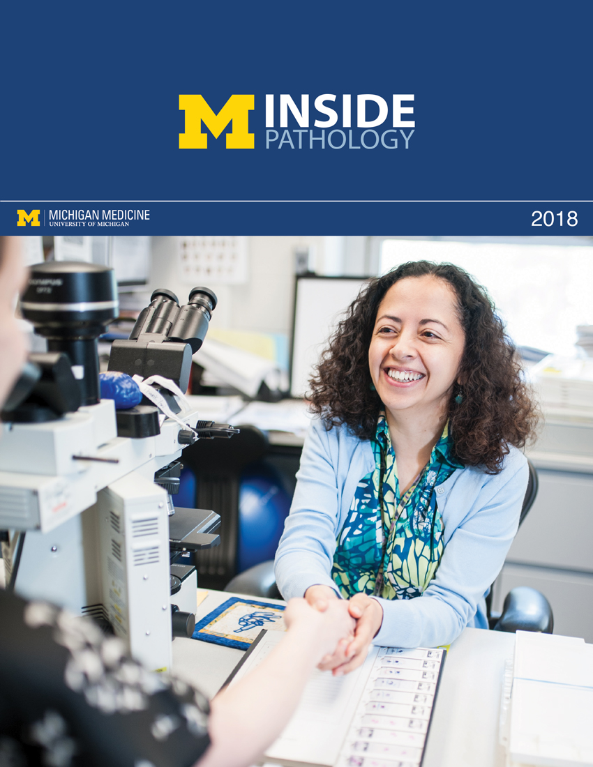 ON THE COVER
Director of the Neuropathology Fellowship, Dr. Sandra Camelo-Piragua serves on the Patient and Family Advisory Council. 2018Department Chair |

newsletter
INSIDE PATHOLOGYAbout Our NewsletterInside Pathology is an newsletter published by the Chairman's Office to bring news and updates from inside the department's research and to become familiar with those leading it. It is our hope that those who read it will enjoy hearing about those new and familiar, and perhaps help in furthering our research. CONTENTS
|
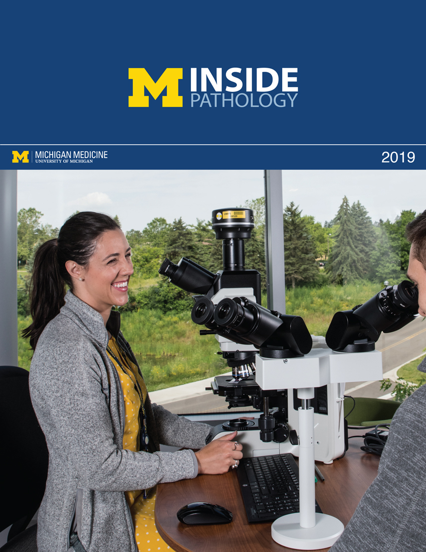 ON THE COVER
Residents Ashley Bradt (left) and William Perry work at a multi-headed scope in our new facility. 2019Department Chair |

newsletter
INSIDE PATHOLOGYAbout Our NewsletterInside Pathology is an newsletter published by the Chairman's Office to bring news and updates from inside the department's research and to become familiar with those leading it. It is our hope that those who read it will enjoy hearing about those new and familiar, and perhaps help in furthering our research. CONTENTS
|
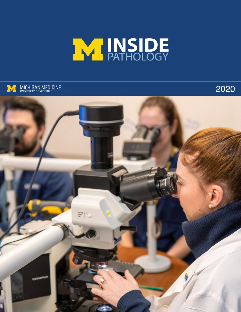 ON THE COVER
Dr. Kristine Konopka (right) instructing residents while using a multi-headed microscope. 2020Department Chair |

newsletter
INSIDE PATHOLOGYAbout Our NewsletterInside Pathology is an newsletter published by the Chairman's Office to bring news and updates from inside the department's research and to become familiar with those leading it. It is our hope that those who read it will enjoy hearing about those new and familiar, and perhaps help in furthering our research. CONTENTS
|
 ON THE COVER
Patient specimens poised for COVID-19 PCR testing. 2021Department Chair |

newsletter
INSIDE PATHOLOGYAbout Our NewsletterInside Pathology is an newsletter published by the Chairman's Office to bring news and updates from inside the department's research and to become familiar with those leading it. It is our hope that those who read it will enjoy hearing about those new and familiar, and perhaps help in furthering our research. CONTENTS
|
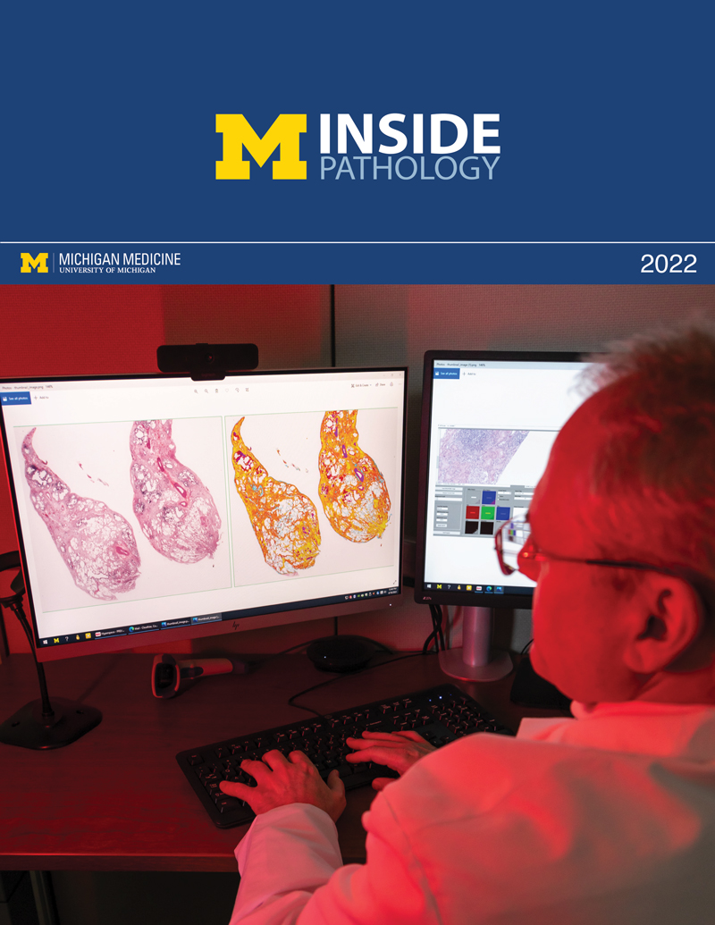 ON THE COVER
Dr. Pantanowitz demonstrates using machine learning in analyzing slides. 2022Department Chair |

newsletter
INSIDE PATHOLOGYAbout Our NewsletterInside Pathology is an newsletter published by the Chairman's Office to bring news and updates from inside the department's research and to become familiar with those leading it. It is our hope that those who read it will enjoy hearing about those new and familiar, and perhaps help in furthering our research. CONTENTS
|
 ON THE COVER
(Left to Right) Drs. Angela Wu, Laura Lamps, and Maria Westerhoff. 2023Department Chair |

newsletter
INSIDE PATHOLOGYAbout Our NewsletterInside Pathology is an newsletter published by the Chairman's Office to bring news and updates from inside the department's research and to become familiar with those leading it. It is our hope that those who read it will enjoy hearing about those new and familiar, and perhaps help in furthering our research. CONTENTS
|
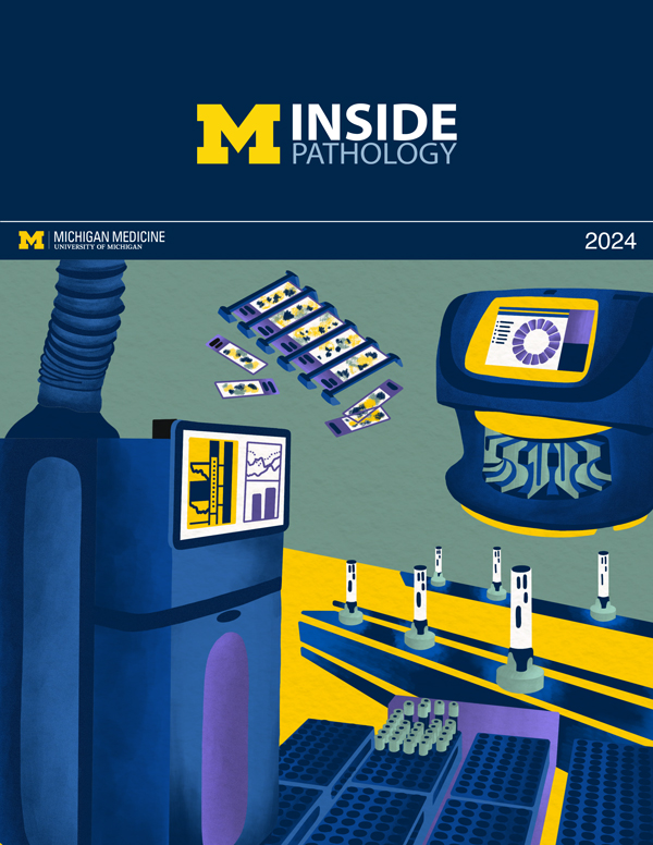 ON THE COVER
Illustration representing the various machines and processing used within our labs. 2024Department Chair |

newsletter
INSIDE PATHOLOGYAbout Our NewsletterInside Pathology is an newsletter published by the Chairman's Office to bring news and updates from inside the department's research and to become familiar with those leading it. It is our hope that those who read it will enjoy hearing about those new and familiar, and perhaps help in furthering our research. CONTENTS
|
 ON THE COVER
Rendering of the D. Dan and Betty Khn Health Care Pavilion. Credit: HOK 2025Department Chair |

newsletter
INSIDE PATHOLOGYAbout Our NewsletterInside Pathology is an newsletter published by the Chairman's Office to bring news and updates from inside the department's research and to become familiar with those leading it. It is our hope that those who read it will enjoy hearing about those new and familiar, and perhaps help in furthering our research. CONTENTS
|

MLabs, established in 1985, functions as a portal to provide pathologists, hospitals. and other reference laboratories access to the faculty, staff and laboratories of the University of Michigan Health System’s Department of Pathology. MLabs is a recognized leader for advanced molecular diagnostic testing, helpful consultants and exceptional customer service.