

Cytogenetics LaboratoryDirector: Lina Shao, MD, PhD, FACMG Oncology Supervisor: Turquessa Brown-Krajewski, B.S., CG(ASCP)CM North Campus Research Building
2800 Plymouth Rd. Bldg. 35
Ann Arbor, MI 48109-2800
(734)763-5805
|
|
|
|
|
||
Services:The UMHS Clinical Cytogenetics Laboratory offers comprehensive cytogenetic testing including standard chromosome analysis (karyotyping), fluorescence in situ hybridization (FISH), and cancer cytogenomic array analysis using whole-genome SNP array. Cytogenetic studies encompass inherited or constitutional disorders such as birth defects, abnormal sexual development, and infertility, as well as neoplasias which are mostly hematologic malignancies, but also some solid tumors. For constitutional studies, many types of specimens are analyzed, including amniotic fluid, chorionic villus samples, tissue biopsies, products of conception, and peripheral blood. A standard peripheral blood constitutional analysis consists of an examination of 20 Giemsa trypsin banded (G-banding) metaphase cells. Every chromosome pair is microscopically analyzed band for band at the 550 band level of resolution, where possible, and at least two karyotypes are prepared. When chromosome analysis is requested to rule out certain conditions, such as Turner syndrome or suspected mosaicism, an additional cell count and/or special stains will be performed. FISH testing is available as an adjunct to chromosome analysis for a wide range of microdeletion syndromes, such as DiGeorge, Prader-Willi, Angelman, Smith-Magenis, Miller-Dieker, and Williams Syndromes. FISH is used to clarify chromosomal rearrangements and identify the origin of marker chromosomes. It is also used to clarify duplications identified by constitutional microarray testing. For chromosome analysis of oncology specimens, which can include bone marrow, lymph nodes or tumor biopsies, at least 20 metaphase cells are analyzed. Over 20 FISH assays are available for assisting diagnosis and classification of malignant hematologic disorders and certain solid tumors, evaluating prognosis and monitoring remission status. For example, identification and monitoring of the BCR/ABL fusion gene in patients with chronic myelogenous leukemia or acute lymphocytic leukemia, for a new diagnosis of APL using a probe for PML/RARA, and for detecting the cryptic t(12;21) in pediatric ALL. The following FISH oncology probes are available: BCR/ABL [t(9;22)], PML/RARA [t(15;17)], RUNX1/RUNX1T1 [t(8;21)], CBFB [inv(16)/t(16;16)], MLL (11q23), MYC (8q24), IGH/CCND1 [t(11;14)], ETV6/RUNX1 [t(12;21)], FIP1L1/PDGFRA [del(4q12)], PDGFRB (5q32), FGFR1 (8p11), and CLL panel. Additional probes may also be available as clinically indicated; contact MLabs or the Cytogenetics laboratory for additional information. FFPE FISH assays for TFE3 (Xp11.2), TFEB (6p21) and ERG (21q22) are also available for classification or prognostication of kidney or prostate malignancies. Cancer cytogenomic array analysis using Affymetrix CytoScan platform is available as complementary for chromosome analysis in myeloid malignancies with a normal karyotype. It is also recommended for all acute lymphoblast leukemia/lymphoma at diagnosis and solid tumors. The microarray detects copy number aberrations and copy neutral loss of heterozygosity. It detects small copy number aberrations that will not be detected by chromosome analysis, such as the IKZF1 deletion in ALL, MLL tandem duplication in AML, and KIAA1497-BRAF tandem duplication in pilocytic astrocytoma. Although cancer cytogenomic array is a powerful diagnostic tool for the evaluation of chromosomal copy number changes, this assay will not detect balanced chromosomal aberrations, imbalance of regions not represented on the microarray, or point mutations. A karyotype or FISH test is more appropriate when a translocation or inversion [e.g., t(8;21), t(9;22), t(15;17), inv(16)] is suspected. Also, chromosome or FISH analysis is more appropriate if a STAT result is required. The detection threshold for mosaicism is variable (20-30%), depending on the size of the segment and tissue type. The Cytogenetics Laboratory processes greater than 4,000 specimens a year and is staffed by over 20 technologists with extensive experience in cytogenetic analysis. The directors are certified in Clinical Genetics by the American Board of Medical Genetics (ABMG). The laboratory is accredited by the College of American Pathologists (CAP) and is an approved lab for Southwest Oncology Group and Children’s Oncology Group (at this time, only University of Michigan registered patient samples are accepted). |
||
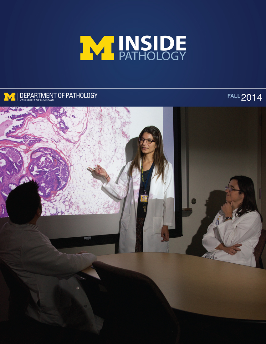 ON THE COVER
Breast team reviewing a patient's slide. (From left to right) Ghassan Allo, Fellow; Laura Walters, Clinical Lecturer; Celina Kleer, Professor. See Article 2014Department Chair |

newsletter
INSIDE PATHOLOGYAbout Our NewsletterInside Pathology is an newsletter published by the Chairman's Office to bring news and updates from inside the department's research and to become familiar with those leading it. It is our hope that those who read it will enjoy hearing about those new and familiar, and perhaps help in furthering our research. CONTENTS
|
 ON THE COVER
Autopsy Technician draws blood while working in the Wayne County morgue. See Article 2016Department Chair |

newsletter
INSIDE PATHOLOGYAbout Our NewsletterInside Pathology is an newsletter published by the Chairman's Office to bring news and updates from inside the department's research and to become familiar with those leading it. It is our hope that those who read it will enjoy hearing about those new and familiar, and perhaps help in furthering our research. CONTENTS
|
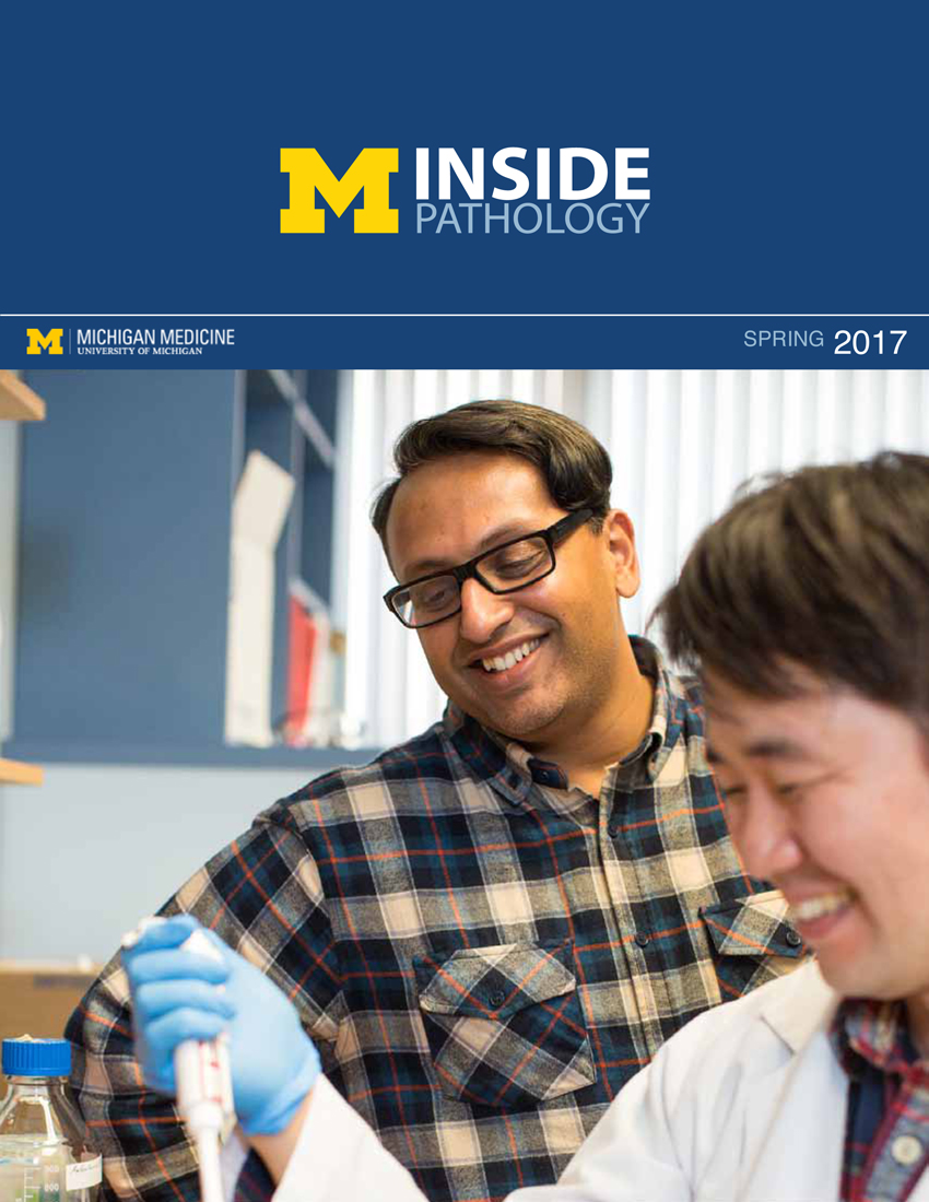 ON THE COVER
Dr. Sriram Venneti, MD, PhD and Postdoctoral Fellow, Chan Chung, PhD investigate pediatric brain cancer. See Article 2017Department Chair |

newsletter
INSIDE PATHOLOGYAbout Our NewsletterInside Pathology is an newsletter published by the Chairman's Office to bring news and updates from inside the department's research and to become familiar with those leading it. It is our hope that those who read it will enjoy hearing about those new and familiar, and perhaps help in furthering our research. CONTENTS
|
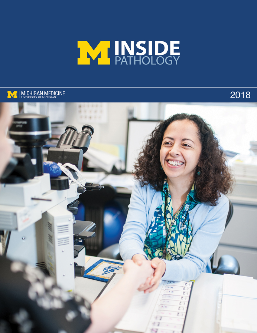 ON THE COVER
Director of the Neuropathology Fellowship, Dr. Sandra Camelo-Piragua serves on the Patient and Family Advisory Council. 2018Department Chair |

newsletter
INSIDE PATHOLOGYAbout Our NewsletterInside Pathology is an newsletter published by the Chairman's Office to bring news and updates from inside the department's research and to become familiar with those leading it. It is our hope that those who read it will enjoy hearing about those new and familiar, and perhaps help in furthering our research. CONTENTS
|
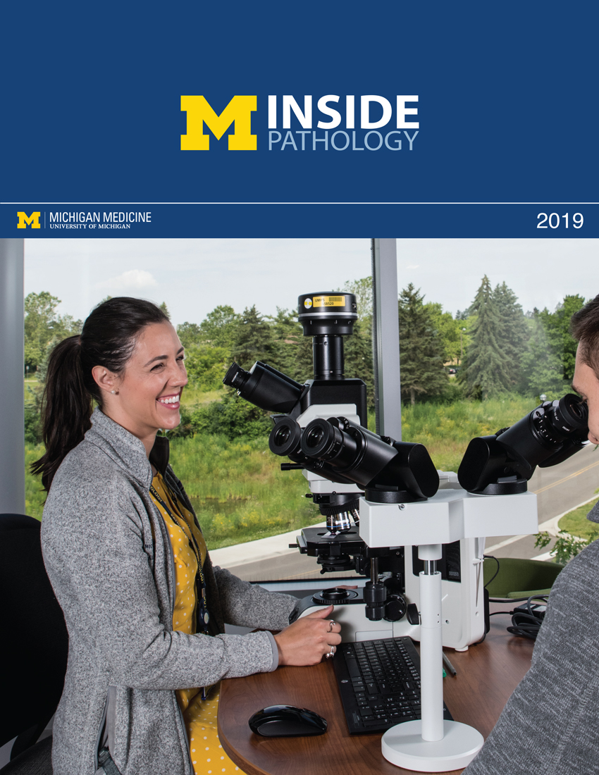 ON THE COVER
Residents Ashley Bradt (left) and William Perry work at a multi-headed scope in our new facility. 2019Department Chair |

newsletter
INSIDE PATHOLOGYAbout Our NewsletterInside Pathology is an newsletter published by the Chairman's Office to bring news and updates from inside the department's research and to become familiar with those leading it. It is our hope that those who read it will enjoy hearing about those new and familiar, and perhaps help in furthering our research. CONTENTS
|
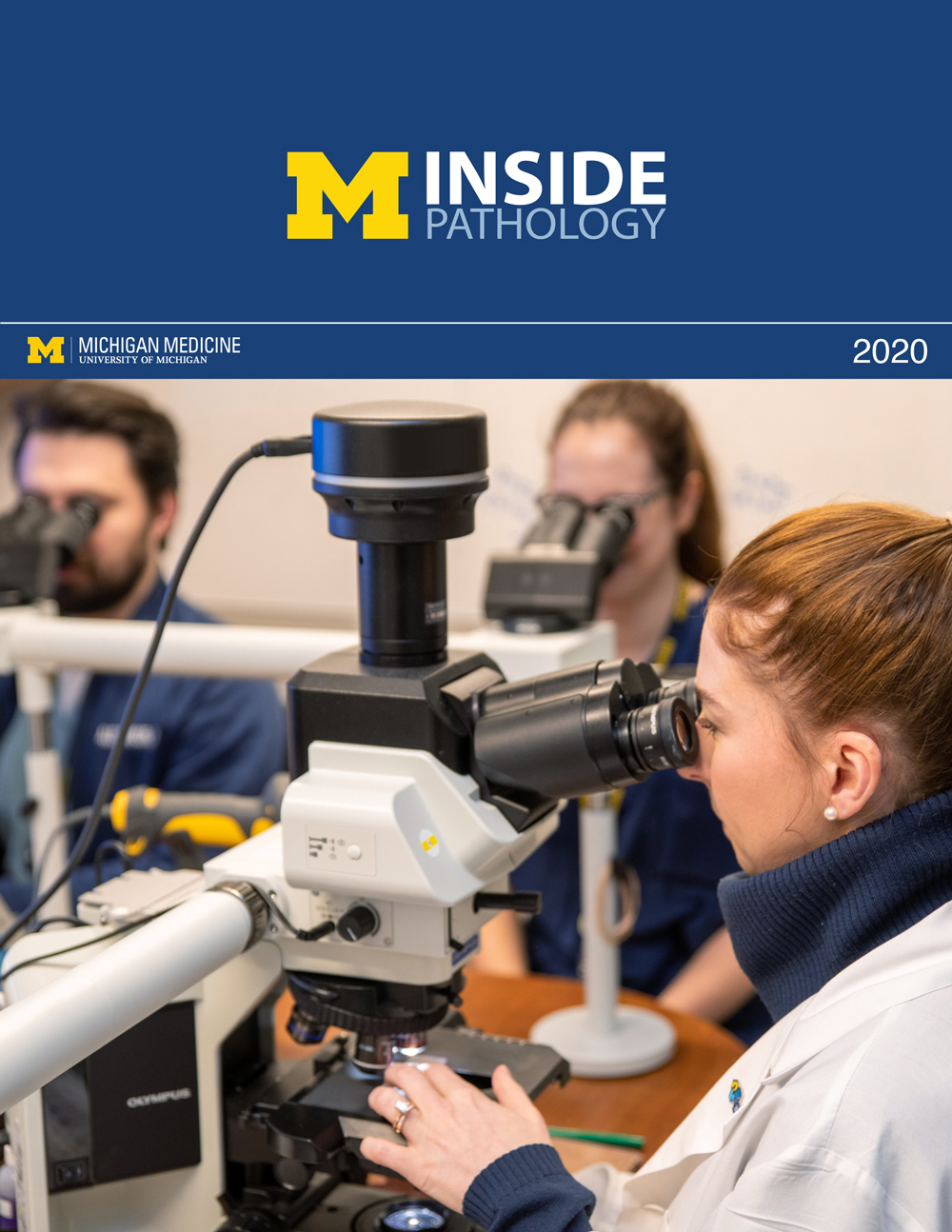 ON THE COVER
Dr. Kristine Konopka (right) instructing residents while using a multi-headed microscope. 2020Department Chair |

newsletter
INSIDE PATHOLOGYAbout Our NewsletterInside Pathology is an newsletter published by the Chairman's Office to bring news and updates from inside the department's research and to become familiar with those leading it. It is our hope that those who read it will enjoy hearing about those new and familiar, and perhaps help in furthering our research. CONTENTS
|
 ON THE COVER
Patient specimens poised for COVID-19 PCR testing. 2021Department Chair |

newsletter
INSIDE PATHOLOGYAbout Our NewsletterInside Pathology is an newsletter published by the Chairman's Office to bring news and updates from inside the department's research and to become familiar with those leading it. It is our hope that those who read it will enjoy hearing about those new and familiar, and perhaps help in furthering our research. CONTENTS
|
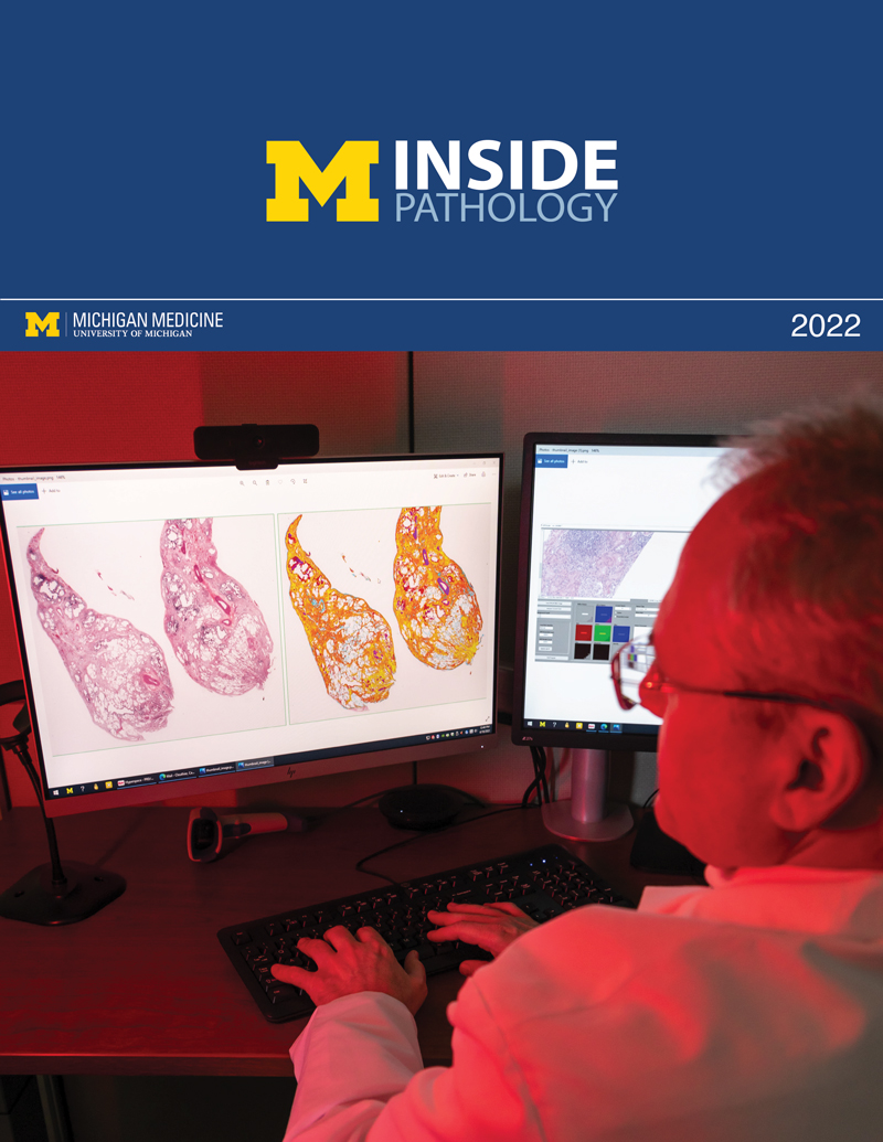 ON THE COVER
Dr. Pantanowitz demonstrates using machine learning in analyzing slides. 2022Department Chair |

newsletter
INSIDE PATHOLOGYAbout Our NewsletterInside Pathology is an newsletter published by the Chairman's Office to bring news and updates from inside the department's research and to become familiar with those leading it. It is our hope that those who read it will enjoy hearing about those new and familiar, and perhaps help in furthering our research. CONTENTS
|
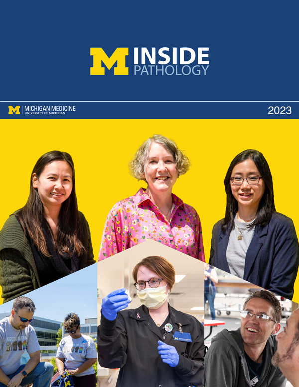 ON THE COVER
(Left to Right) Drs. Angela Wu, Laura Lamps, and Maria Westerhoff. 2023Department Chair |

newsletter
INSIDE PATHOLOGYAbout Our NewsletterInside Pathology is an newsletter published by the Chairman's Office to bring news and updates from inside the department's research and to become familiar with those leading it. It is our hope that those who read it will enjoy hearing about those new and familiar, and perhaps help in furthering our research. CONTENTS
|
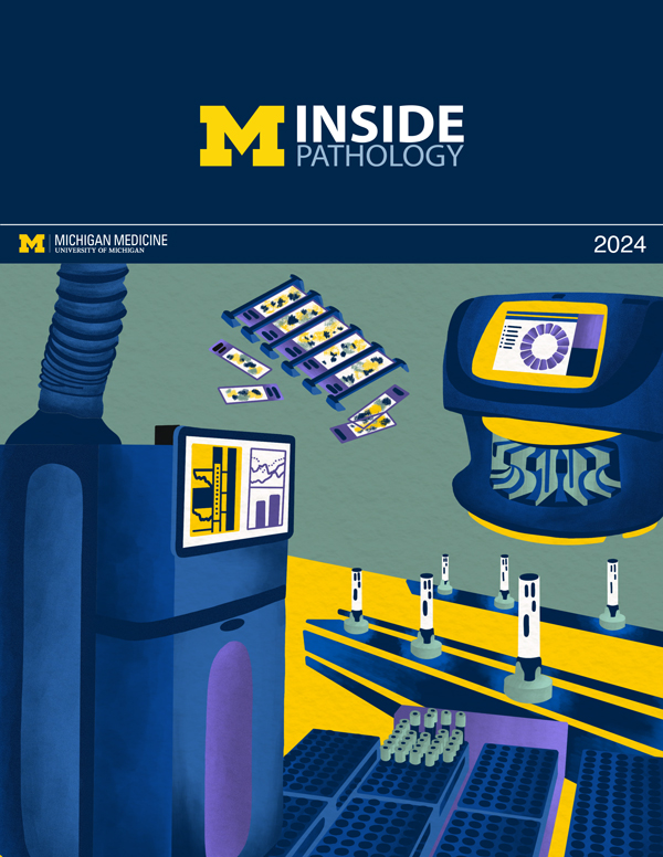 ON THE COVER
Illustration representing the various machines and processing used within our labs. 2024Department Chair |

newsletter
INSIDE PATHOLOGYAbout Our NewsletterInside Pathology is an newsletter published by the Chairman's Office to bring news and updates from inside the department's research and to become familiar with those leading it. It is our hope that those who read it will enjoy hearing about those new and familiar, and perhaps help in furthering our research. CONTENTS
|
 ON THE COVER
Rendering of the D. Dan and Betty Khn Health Care Pavilion. Credit: HOK 2025Department Chair |

newsletter
INSIDE PATHOLOGYAbout Our NewsletterInside Pathology is an newsletter published by the Chairman's Office to bring news and updates from inside the department's research and to become familiar with those leading it. It is our hope that those who read it will enjoy hearing about those new and familiar, and perhaps help in furthering our research. CONTENTS
|

MLabs, established in 1985, functions as a portal to provide pathologists, hospitals. and other reference laboratories access to the faculty, staff and laboratories of the University of Michigan Health System’s Department of Pathology. MLabs is a recognized leader for advanced molecular diagnostic testing, helpful consultants and exceptional customer service.