
5 Blood Transfusion Guidelines and Utilization Review
Transfusion decisions should be based on clinical assessment of the patient and laboratory test results. There are no absolute indications and few contraindications to blood transfusion. These guidelines are intended as an aid in decision making.
Red Blood Cells
Red blood cells (RBC) are indicated for increasing the oxygen carrying capacity in anemia patients. In addition, RBC transfusion can increase intravascular volume and improve platelet function, particularly in uremic patients. Considerations in ordering RBC transfusion include:
|
Weight Kg |
estimated blood volume |
pre hematocrit |
post hematocrit |
percent of red cell mass lost |
estimated blood volume lost |
|
60 |
4200 |
35 |
30 |
14% |
600 |
|
3500 |
30 |
25 |
17% |
583 |
|
|
3500 |
25 |
20 |
20% |
700 |
|
|
70 |
4900 |
35 |
30 |
14% |
700 |
|
4900 |
30 |
25 |
17% |
817 |
|
|
4900 |
25 |
20 |
20% |
980 |
|
|
80 |
5600 |
35 |
30 |
14% |
800 |
|
5600 |
30 |
25 |
17% |
933 |
|
|
5600 |
25 |
20 |
20% |
1120 |
General indications for RBC transfusion include:
RBC dosage
One unit of RBC can be expected to result in a hemoglobin increase of 1 g/dl or hematocrit increase of 3% in a typical adult. One unit of RBC can replace a blood loss of 500 ml.
Monitoring RBC transfusion effectiveness
The patient should be assessed and post-transfusion hemoglobin measured to monitor the effectiveness of transfusion. Lack of clinical benefit may indicate ongoing blood loss or cardiac or pulmonary disease. The Blood Bank house officer should be consulted if a patient has an inadequate response to transfusion and other causes are excluded. Causes of a less than expected hemoglobin response include:
Platelets
Platelets are indicated for the prevention or control of bleeding due to thrombocytopenia or platelet dysfunction. Platelets may be provided as pooled whole-blood derived platelet concentrates ("random donor" platelets) and as apheresis platelet concentrates ("single donor" platelets). For most patients these products are equally effective. Apheresis platelets are indicated for patients with immune refractoriness when crossmatched or HLA matched platelets have better post-transfusion survival.
Considerations in ordering platelet transfusions include:
General indications for platelet transfusion include:
Platelet dosage
Transfusion of one platelet pool and one unit of apheresis platelets will typically increase the platelet count of an adult by 20,000 – 40,000/microL. Alternatively, transfusion of one unit per 10 kg body weight will typically increase the platelet count by 10,000/l. Platelets should be transfused immediately before or during invasive procedure for maximally effectiveness. In a patient with normal splenic function, approximately 40% of transfused platelets will be sequestered in the spleen. This proportion is increased in splenomegaly.
Monitoring platelet transfusion effectiveness
A post-transfusion platelet count should be obtained 10 minutes to 1 hour after transfusion for best assessment of transfusion effectiveness. Platelet counts obtained later may not allow for differentiation between immune and non-immune causes of platelet transfusion refractoriness. The corrected count increment (CCI) is usually the best assessment of transfusion effectiveness. A one-hour CCI greater than 5,000 is typically considered a satisfactory response. The Blood Bank house officer should be consulted when platelet transfusion refractoriness is suspected.
Causes of an inadequate response to platelet transfusion include:
|
Heparin |
Fibrinolytic agents |
|
Platelet function inhibitors |
Nitroglycerin, insosorbide, nitroprusside |
|
Non-steroidal anti-inflammatory drugs |
Beta blockers |
|
Antibiotics including beta lactams, nitrofurantions, and vancomycin |
Calcium channel blockers |
|
Antifungals, particularly amphotericin |
Quinidine |
|
Dipyriamole |
Phenothiazines and tricyclic antidepressants |
|
Local and general anesthetic agents |
Radiographic contrast agents |
|
Biological such as anti-thymocyte globulin and abciximab |
|
If refractoriness is suspected, the Blood Bank house officer should be consulted. Generally, three one-hour post-transfusion platelet counts with poor increments are necessary to establish refractoriness. Blood samples for platelet crossmatch should be sent to the Blood Bank and for HLA antibodiesshould be set to the Tissue Typing laboratory. The determination of immune refractoriness and selection of platelet components will depend on these results. Additional studies for platelet specific antibodies or HLA typing of the recipient may be requested by the Blood Bank.
Contraindication to platelet transfusion
Platelet transfusion is contraindicated in thrombotic thrombocytopenic purpura (TTP) and heparin-induced thrombocytopenic (HIT). Serious adverse events have occurred with platelet transfusion in these settings. Platelet transfusion is relatively contraindicated in immune thrombocytopenic purpura (ITP) or post-transfusion purpura (PTP) because the survival of transfused platelets is extremely brief.
Fresh Frozen Plasma
Plasma is provided as Fresh Frozen plasma (FFP) or Liquid Plasma ("single donor" plasma). FFP is plasma within 24 hours of thawing. After 24 hours, thawed plasma may be relabeled as liquid plasma and stored at 4°C for up to 5 days. Liquid Plasma has essentially the same coagulation factor content as FFP and may be used interchangeably for most patients. Plasma may be transfused for replacement of any plasma protein deficiency, usually coagulation factor deficiency.
General indications for plasma transfusion include:
Plasma dosage
A dose of 10 ml/kg will typically provide sufficient coagulation factors to achieve hemostasis. Factor levels in donor plasma are variable, but can be assumed to be approximately 1 U/ml. Post-transfusion recovery of transfused factors may be less than expected due to extravascular distribution or consumption.
The following table may be useful as a guide to coagulation factor replacement.
|
Factor |
Plasma Concentration Required for Hemostasis (U/ml) |
Half-Life of Transfused Factor |
Recovery in Blood (as % of Amount Transfused) |
|
I (fibrinogen) |
100-150 mg/dL |
3-6 days |
50% |
|
II |
0.4 |
2-5 days |
40-80% |
|
V |
0.1 – 0.25 |
15-36 hours |
80% |
|
VII |
0.05 – 0.2 |
2-7 hours |
70-80% |
|
VIII |
0.1 – 0.40 |
8-12 hours |
60-80% |
|
IX |
0.1 – 0.4 |
18-24 hours |
40-50% |
|
X |
0.1 – 0.2 |
1.5-2 days |
50% |
|
XI |
0.15 – 0.3 |
3-4 days |
90-100% |
|
XIII |
0.1 – 0.5 |
6-10 days |
5-100% |
|
vWF |
0.25 – 0.5 |
3-5 hours |
|
Monitoring plasma transfusion effectiveness
Plasma transfusion should be monitored with specific factor levels or PT and aPTT measurements within 4 hours of transfusion. Causes of an inadequate response to plasma transfusion include:
Cryoprecipitate
Cryoprecipitated antihemophilic factor (CAF or "cryo") is a concentrate of Factor VIII, von Willebrand’s factor, fibrinogen, and Factor XIII. Each unit contains a minimum of 80 U of Factor VIII and typically 250 mg of fibrinogen. Cryoprecipitate is generally indicated for:
Cryoprecipitate is not a significant source of other coagulation factors, and cannot be used as an alternative to plasma.
Cryoprecipitate dosage
For Factor VIII replacement, the dose can be calculated assuming 80 U per bag. Current methods of manufacturing typically yield higher Factor VIII content. Contact the Blood Bank for current information of Factor VIII levels. For fibrinogen replacement, the dose can be calculated assuming 250 mg per bag. For other indications, cryoprecipitate is usually given as 1 unit per 10 kg, or a pool of 10 units for an adult. For topical fibrin adhesive, typically 25 – 50 ml are ordered.
Monitoring cryoprecipitate transfusion
Clinical response is usually the best assessment of cryoprecipitate transfusion effectiveness. Factor VIII activity, fibrinogen, or von Willebrand’s factor activity should be measured 1 hour after transfusion. Lack of expected benefit may be due to:
Massive Transfusion
Massive transfusion is defined as the replacement of one blood volume within 24 hours. This is approximately equivalent to transfusion of 10 units of Red Blood Cells in an adult. Coagulopathy due to thrombocytopenia, factor consumption, or factor dilution may occur in massive transfusion. Optimal management should be based on frequent clinical assessment and laboratory monitoring. Consideration should be paid to:
Patient assessment should include:
Assessments should be repeated frequently (typically after each 5-10 units of red cells transfused). No single transfusion protocol is applicable for all massively transfused patients, but general guidelines include:
Mediastinal bleeding following cardiac surgery
Excessive bleeding after cardiac surgery may be due to heparin, coagulation factor deficiency, thrombocytopenia, surgical factors, or underlying coagulopathy. Evaluation of the postoperative patient should include:
Surgical bleeding is generally indicated by:
An elevated heparin level, typically with prolongation of the aPTT, indicates that unneutralized heparin is present, in which case protamine is generally indicated. Coagulopathy may be indicated by:
A low fibrinogen may be due to fibrinolysis, in which case -aminocaproic acid may be indicated. Elevated PT or aPTT after heparin neutralization is usually an indication for plasma transfusion, 10 ml/kg. Microvascular bleeding or elevated chest tube output with platelet count < 50,000/l is usually an indication for transfusion of 1 platelet pool or 1 apheresis platelet unit.
Liver disease
Factors contributing to bleeding in liver disease include:
Utilization Review
Blood transfusion practices at the University of Michigan Hospitals and Health Centers are reviewed by the Transfusion Committee of the Medical Staff. The purpose of a utilization review is to improve the processes involved in the ordering, distribution, handling, dispensing and administration of blood components and to monitor the effects of transfusion practices. Conducting such audits is an accreditation requirement of the Joint Commission on the Accreditation of Healthcare Organizations (JCAHO).
The review criteria are approved by the Transfusion Committee, and the chairs of the clinical departments. These criteria reflect a consensus as to the generally accepted rationale for the use of blood components based published clinical trials, consensus statements, and guidelines produced by national organizations. However, it must be noted that review criteria do not necessarily constitute indications, or triggers, for transfusion and that specific clinical situations may dictate transfusion practices that differ from the review criteria. The Transfusion Committee recognizes that all transfusion decisions are clinical judgments that cannot necessarily be reduced to predefined indications.
Utilization reviews are generally focused on procedures and patient care units with high use, patients requiring special products, or transfusion situations at increased risk of adverse outcomes. Elements of utilization review include:
References
Version July 2004
 ON THE COVER
Breast team reviewing a patient's slide. (From left to right) Ghassan Allo, Fellow; Laura Walters, Clinical Lecturer; Celina Kleer, Professor. See Article |
newsletter
INSIDE PATHOLOGYAbout Our NewsletterInside Pathology is an newsletter published by the Chairman's Office to bring news and updates from inside the department's research and to become familiar with those leading it. It is our hope that those who read it will enjoy hearing about those new and familiar, and perhaps help in furthering our research. CONTENTS
|
 ON THE COVER
Autopsy Technician draws blood while working in the Wayne County morgue. See Article |
newsletter
INSIDE PATHOLOGYAbout Our NewsletterInside Pathology is an newsletter published by the Chairman's Office to bring news and updates from inside the department's research and to become familiar with those leading it. It is our hope that those who read it will enjoy hearing about those new and familiar, and perhaps help in furthering our research. CONTENTS
|
 ON THE COVER
Dr. Sriram Venneti, MD, PhD and Postdoctoral Fellow, Chan Chung, PhD investigate pediatric brain cancer. See Article |
newsletter
INSIDE PATHOLOGYAbout Our NewsletterInside Pathology is an newsletter published by the Chairman's Office to bring news and updates from inside the department's research and to become familiar with those leading it. It is our hope that those who read it will enjoy hearing about those new and familiar, and perhaps help in furthering our research. CONTENTS
|
 ON THE COVER
Director of the Neuropathology Fellowship, Dr. Sandra Camelo-Piragua serves on the Patient and Family Advisory Council. |
newsletter
INSIDE PATHOLOGYAbout Our NewsletterInside Pathology is an newsletter published by the Chairman's Office to bring news and updates from inside the department's research and to become familiar with those leading it. It is our hope that those who read it will enjoy hearing about those new and familiar, and perhaps help in furthering our research. CONTENTS
|
 ON THE COVER
Residents Ashley Bradt (left) and William Perry work at a multi-headed scope in our new facility. |
newsletter
INSIDE PATHOLOGYAbout Our NewsletterInside Pathology is an newsletter published by the Chairman's Office to bring news and updates from inside the department's research and to become familiar with those leading it. It is our hope that those who read it will enjoy hearing about those new and familiar, and perhaps help in furthering our research. CONTENTS
|
 ON THE COVER
Dr. Kristine Konopka (right) instructing residents while using a multi-headed microscope. |
newsletter
INSIDE PATHOLOGYAbout Our NewsletterInside Pathology is an newsletter published by the Chairman's Office to bring news and updates from inside the department's research and to become familiar with those leading it. It is our hope that those who read it will enjoy hearing about those new and familiar, and perhaps help in furthering our research. CONTENTS
|
 ON THE COVER
Patient specimens poised for COVID-19 PCR testing. |
newsletter
INSIDE PATHOLOGYAbout Our NewsletterInside Pathology is an newsletter published by the Chairman's Office to bring news and updates from inside the department's research and to become familiar with those leading it. It is our hope that those who read it will enjoy hearing about those new and familiar, and perhaps help in furthering our research. CONTENTS
|
 ON THE COVER
Dr. Pantanowitz demonstrates using machine learning in analyzing slides. |
newsletter
INSIDE PATHOLOGYAbout Our NewsletterInside Pathology is an newsletter published by the Chairman's Office to bring news and updates from inside the department's research and to become familiar with those leading it. It is our hope that those who read it will enjoy hearing about those new and familiar, and perhaps help in furthering our research. CONTENTS
|
 ON THE COVER
(Left to Right) Drs. Angela Wu, Laura Lamps, and Maria Westerhoff. |
newsletter
INSIDE PATHOLOGYAbout Our NewsletterInside Pathology is an newsletter published by the Chairman's Office to bring news and updates from inside the department's research and to become familiar with those leading it. It is our hope that those who read it will enjoy hearing about those new and familiar, and perhaps help in furthering our research. CONTENTS
|
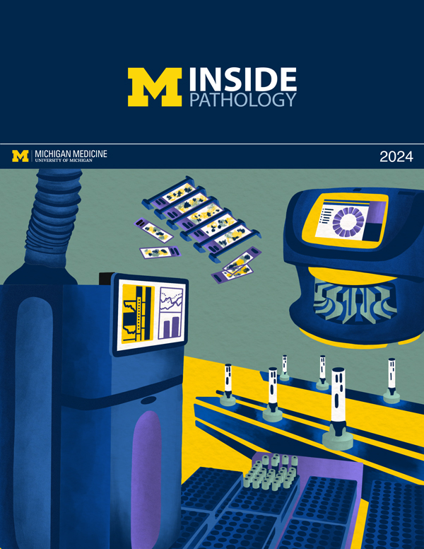 ON THE COVER
Illustration representing the various machines and processing used within our labs. |
newsletter
INSIDE PATHOLOGYAbout Our NewsletterInside Pathology is an newsletter published by the Chairman's Office to bring news and updates from inside the department's research and to become familiar with those leading it. It is our hope that those who read it will enjoy hearing about those new and familiar, and perhaps help in furthering our research. CONTENTS
|
 ON THE COVER
Rendering of the D. Dan and Betty Khn Health Care Pavilion. Credit: HOK |
newsletter
INSIDE PATHOLOGYAbout Our NewsletterInside Pathology is an newsletter published by the Chairman's Office to bring news and updates from inside the department's research and to become familiar with those leading it. It is our hope that those who read it will enjoy hearing about those new and familiar, and perhaps help in furthering our research. CONTENTS
|
MLabs, established in 1985, functions as a portal to provide pathologists, hospitals. and other reference laboratories access to the faculty, staff and laboratories of the University of Michigan Health System’s Department of Pathology. MLabs is a recognized leader for advanced molecular diagnostic testing, helpful consultants and exceptional customer service.