

By Beth Uznis Johnson | healthblog.uofmhealth.org | 24 January
 Chances are, the treatment plan for your cancer was determined by the results presented on a pathology report.
This process probably started before your diagnosis, when you had a biopsy or surgery where a doctor removed cells or tissue for study under a microscope. Specialists called pathologists studied these samples to understand how they look compared with normal cells and prepared a report summarizing their findings for your oncologist or surgeon. Patients may not see reports like these, nor understand the technical medical language used.
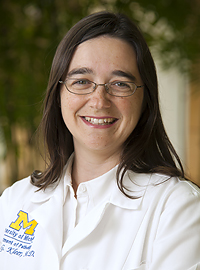
To help patients understand the information in each report and how your oncologist uses it to decide the best course of treatment for your cancer, University of Michigan Comprehensive Cancer Center pathologist Celina Kleer, M.D., director of the breast pathology program, explains more.
Kleer: Each report has patient identifiers, including the name, birthdate, date of procedure and an assigned pathology accession number. The report contains a macroscopic (also called gross) description of the tissue sample. This consists of the features of the tissue or organ seen with the naked eye, before study under the microscope. Under the diagnosis heading is a statement of whether the lesion excised is benign or malignant, and the specific medical term used to categorize the abnormal findings. The pathology report also has a section labeled “comments,” where the pathologist may note special features or unusual aspects of the sample.
Here is an example of a breast biopsy diagnosis:
Gross description: A 3x2x2 cm oval fragment of fibrous and adipose tissue is received fresh and labeled “left breast biopsy.” Upon sectioning, there is a 1 cm tan firm nodule with a glistening surface. The nodule is sectioned and submitted entirely for review. In addition, there are multiple gritty yellow areas surrounding the nodule, measuring approximately 0.5 cm.
Diagnosis: Left breast, excisional biopsy: High nuclear grade ductal carcinoma in situ with microcalcifications surrounding a fibroadenoma. Ductal carcinoma in situ extends to margins. See Comment.
Comment: The foci of ductal carcinoma in situ have focal necrosis and marked nuclear pleomorphism. Hormonal receptors will be performed.
All of this information will be explained to you by your oncologist. Be sure to ask questions if there are medical terms or details you don’t understand.
Kleer: The pathology report states as precisely as possible the disease present in the tissue sample. The aim of the report is to provide the clinicians and surgeons with the necessary information so they can make a treatment recommendation. The pathologist can also perform additional tests on the tissues that can inform on the rate of growth and how the disease may evolve. For example, finding cellular atypia and multiple division figures (or mitosis) may indicate that the tumor is growing rapidly, and guides oncologists and surgeons on the most appropriate treatment course.
Kleer: Once the pathologist writes a report, the physician caring for the patient will communicate the results and explain the meaning of the diagnosis rendered, which will lead to a treatment plan. Pathology reports contain medical and scientific terms that may be difficult for nonmedical people to understand. Thus, it is important to discuss all diagnoses with your primary doctor.
Kleer: If genetic testing is indicated, based on the particular disease, this information is included in a special genetic report. The Cancer Center and the Department of Pathology has uniquely qualified specialists to perform genetic testing. Some patients have their tumors sequenced to find specific mutations that can be targeted with available medicines. This personalized medicine approach requires a team of physicians of different specialties including clinicians and pathologists.
Kleer: I think second opinions are always worthwhile. Here at the U-M Cancer Center, each breast biopsy is reviewed several times. First, a pathology resident reviews the slides. Then, the resident reviews the case with a pathology fellow and an attending pathologist. That’s already three sets of eyes. If there are any doubts about a diagnosis, the case will be reviewed with another pathologist.
It is important to note that a multidisciplinary team (including radiologists, oncologists, pathologists, radiation oncologists, surgeons, nurses, geneticists, psychiatrists and social workers) is closely involved in the decision-making and care of cancer patients. Their close communication is essential in providing optimal treatment.
For example, in breast cancer, here at the Cancer Center, we present each patient’s case to our breast multidisciplinary tumor board. We show slides on a projector and discuss the diagnosis and treatment plan of each individual patient. Everyone is able to offer his or her expertise.
The previously mentioned reviews all take place before this meeting.
Often, smaller hospitals and clinics send cases out for consultation to larger institutions like the University of Michigan. In our pathology department, we evaluate multiple cases from regional hospitals.
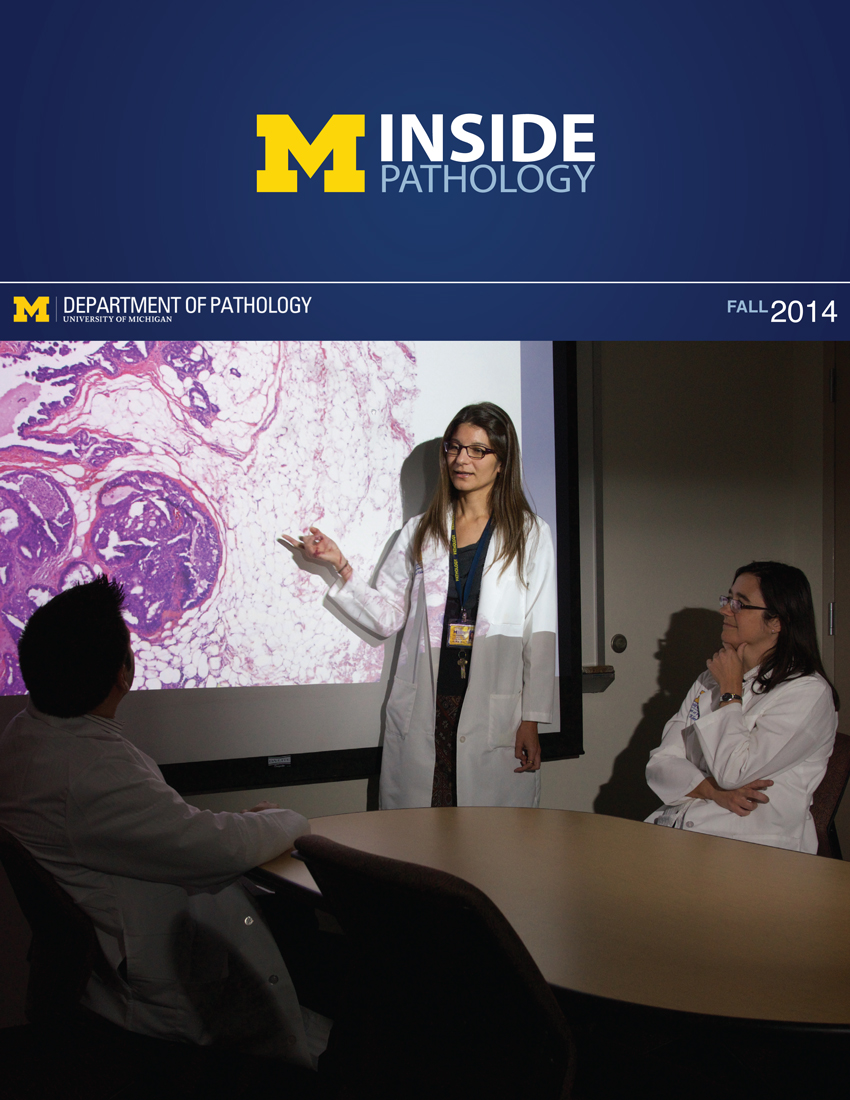 ON THE COVER
Breast team reviewing a patient's slide. (From left to right) Ghassan Allo, Fellow; Laura Walters, Clinical Lecturer; Celina Kleer, Professor. See Article 2014Department Chair |

newsletter
INSIDE PATHOLOGYAbout Our NewsletterInside Pathology is an newsletter published by the Chairman's Office to bring news and updates from inside the department's research and to become familiar with those leading it. It is our hope that those who read it will enjoy hearing about those new and familiar, and perhaps help in furthering our research. CONTENTS
|
 ON THE COVER
Autopsy Technician draws blood while working in the Wayne County morgue. See Article 2016Department Chair |

newsletter
INSIDE PATHOLOGYAbout Our NewsletterInside Pathology is an newsletter published by the Chairman's Office to bring news and updates from inside the department's research and to become familiar with those leading it. It is our hope that those who read it will enjoy hearing about those new and familiar, and perhaps help in furthering our research. CONTENTS
|
 ON THE COVER
Dr. Sriram Venneti, MD, PhD and Postdoctoral Fellow, Chan Chung, PhD investigate pediatric brain cancer. See Article 2017Department Chair |

newsletter
INSIDE PATHOLOGYAbout Our NewsletterInside Pathology is an newsletter published by the Chairman's Office to bring news and updates from inside the department's research and to become familiar with those leading it. It is our hope that those who read it will enjoy hearing about those new and familiar, and perhaps help in furthering our research. CONTENTS
|
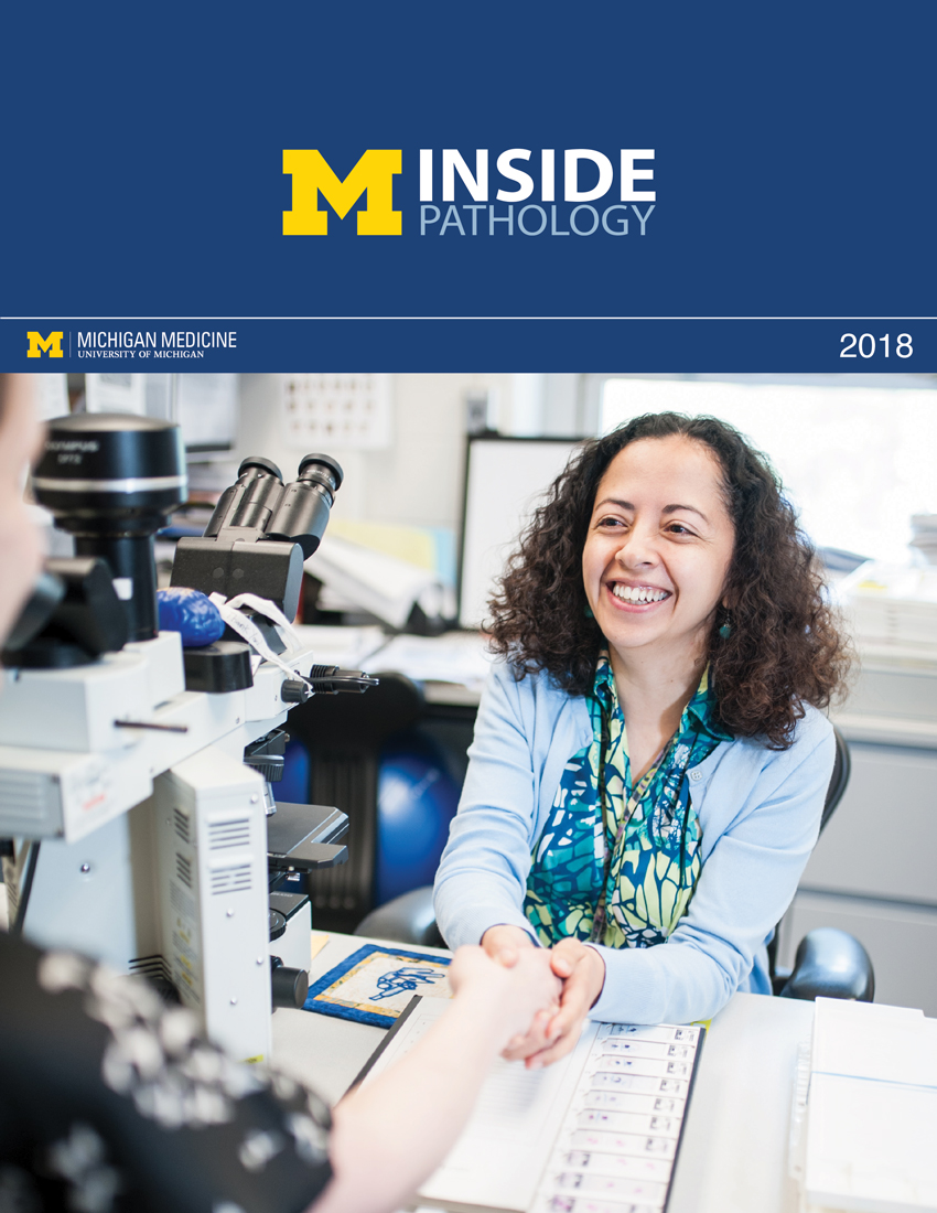 ON THE COVER
Director of the Neuropathology Fellowship, Dr. Sandra Camelo-Piragua serves on the Patient and Family Advisory Council. 2018Department Chair |

newsletter
INSIDE PATHOLOGYAbout Our NewsletterInside Pathology is an newsletter published by the Chairman's Office to bring news and updates from inside the department's research and to become familiar with those leading it. It is our hope that those who read it will enjoy hearing about those new and familiar, and perhaps help in furthering our research. CONTENTS
|
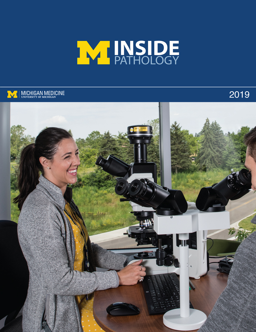 ON THE COVER
Residents Ashley Bradt (left) and William Perry work at a multi-headed scope in our new facility. 2019Department Chair |

newsletter
INSIDE PATHOLOGYAbout Our NewsletterInside Pathology is an newsletter published by the Chairman's Office to bring news and updates from inside the department's research and to become familiar with those leading it. It is our hope that those who read it will enjoy hearing about those new and familiar, and perhaps help in furthering our research. CONTENTS
|
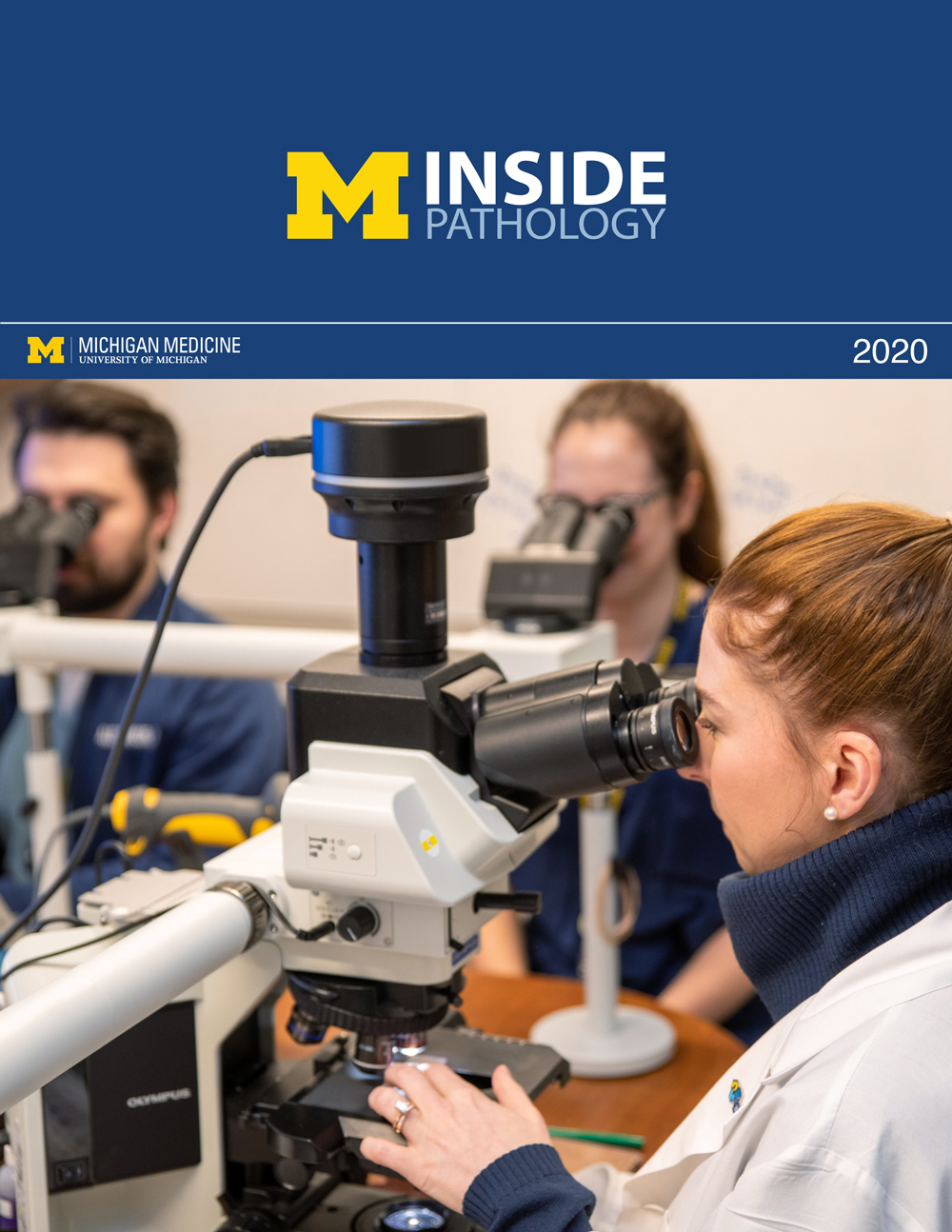 ON THE COVER
Dr. Kristine Konopka (right) instructing residents while using a multi-headed microscope. 2020Department Chair |

newsletter
INSIDE PATHOLOGYAbout Our NewsletterInside Pathology is an newsletter published by the Chairman's Office to bring news and updates from inside the department's research and to become familiar with those leading it. It is our hope that those who read it will enjoy hearing about those new and familiar, and perhaps help in furthering our research. CONTENTS
|
 ON THE COVER
Patient specimens poised for COVID-19 PCR testing. 2021Department Chair |

newsletter
INSIDE PATHOLOGYAbout Our NewsletterInside Pathology is an newsletter published by the Chairman's Office to bring news and updates from inside the department's research and to become familiar with those leading it. It is our hope that those who read it will enjoy hearing about those new and familiar, and perhaps help in furthering our research. CONTENTS
|
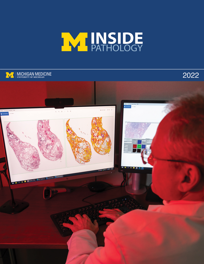 ON THE COVER
Dr. Pantanowitz demonstrates using machine learning in analyzing slides. 2022Department Chair |

newsletter
INSIDE PATHOLOGYAbout Our NewsletterInside Pathology is an newsletter published by the Chairman's Office to bring news and updates from inside the department's research and to become familiar with those leading it. It is our hope that those who read it will enjoy hearing about those new and familiar, and perhaps help in furthering our research. CONTENTS
|
 ON THE COVER
(Left to Right) Drs. Angela Wu, Laura Lamps, and Maria Westerhoff. 2023Department Chair |

newsletter
INSIDE PATHOLOGYAbout Our NewsletterInside Pathology is an newsletter published by the Chairman's Office to bring news and updates from inside the department's research and to become familiar with those leading it. It is our hope that those who read it will enjoy hearing about those new and familiar, and perhaps help in furthering our research. CONTENTS
|
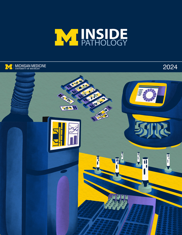 ON THE COVER
Illustration representing the various machines and processing used within our labs. 2024Department Chair |

newsletter
INSIDE PATHOLOGYAbout Our NewsletterInside Pathology is an newsletter published by the Chairman's Office to bring news and updates from inside the department's research and to become familiar with those leading it. It is our hope that those who read it will enjoy hearing about those new and familiar, and perhaps help in furthering our research. CONTENTS
|
 ON THE COVER
Rendering of the D. Dan and Betty Khn Health Care Pavilion. Credit: HOK 2025Department Chair |

newsletter
INSIDE PATHOLOGYAbout Our NewsletterInside Pathology is an newsletter published by the Chairman's Office to bring news and updates from inside the department's research and to become familiar with those leading it. It is our hope that those who read it will enjoy hearing about those new and familiar, and perhaps help in furthering our research. CONTENTS
|

MLabs, established in 1985, functions as a portal to provide pathologists, hospitals. and other reference laboratories access to the faculty, staff and laboratories of the University of Michigan Health System’s Department of Pathology. MLabs is a recognized leader for advanced molecular diagnostic testing, helpful consultants and exceptional customer service.