Digital pathology has the potential to transform routine pathology practice. It is generally defined as the digitization of pathology slides and the management of data associated with them. The Department of Pathology’s digital pathology practice is today part of the Division of Pathology Informatics, headed by Drs. Ulysses Balis and David McClintock.
 Over a decade ago, under the direction of Dr. Lloyd Stoolman, the Department began using scanned slide images, often called whole-slide images, for pathology education. In 2014, Peter Ouillette, MT (ASCP) was hired by the Department to manage routine scanning operations.
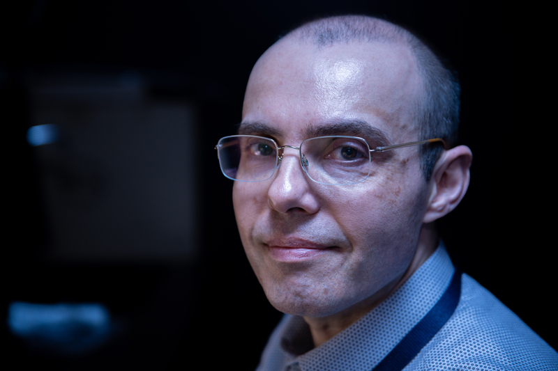
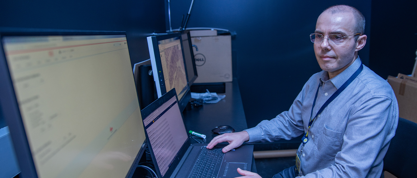
Ouillette began his career as a Medical Technologist at Mayo Clinic in Rochester, Minnesota, where he worked for 7 years before coming to Ann Arbor in 2004 to work in a leukemia research lab. In 2014, he responded to a Pathology Department job posting for someone to scan slides and manage the Department’s research flow cytometers. Today, he is a valuable member of the Pathology Informatics team, and digital pathology is poised to grow significantly in the near future. In five years, he has seen the annual volume of slides scanned rise from 7000 to over 23,000; he has also implemented “telepathology” for frozen section diagnoses. Ouillette sat down with the Pathology Communications team recently to shed light on his work, the processes, and the roles of the expanding and changing technology that is digital pathology.
Q: What is your role here in the Department of Pathology?
A: I am the Operations Manager for Digital Pathology. We primarily deliver high-quality whole-slide images to pathologists, trainees, and researchers in support of tumor board conferences, education, and patient care other than for primary diagnosis. I work with Pathology Informatics programmers to support web-based pathology education resources freely available throughout the world. Finally, I also work to develop, validate, implement, and support telepathology solutions for primary diagnoses.
Q: What is the mission of your team?
A: It’s my goal to see that all of these tasks are performed in a can-do, customer-centered spirit. We digitize glass pathology slides through our slide scanning instruments. If you need to identify the patient whose tissue is represented by that digital slide, plus describe what is important or diagnostic about that tissue, we catalog all of that information, so it is easily accessible. The digital images need to be stored in a manner that is redundant, reliable, capacious enough, and can be grown to handle future needs. We play a critical role in that as well. We act as the technical resource, the liaison, between pathologists and IT solution vendors to help make this happen in a way that fulfills Pathology’s specific goals and use cases.
Q: What is an example of custom work that you have done for a pathologist?
A: A big project that we took on beginning in 2018 was on behalf of Jeffrey Hodgin, MD, PhD; a nephropathologist who also runs a research laboratory here at Michigan Medicine. Jeff’s vision was to establish a shared cohort of kidney biopsy digital images and present it to collaborators around the world. We needed to set up a new server and instance of database software, both of which were on a public IP address so that they would be accessible from outside U-M. To date, we have scanned thousands of de-identified slides in pursuit of this project and stand ready to assist with making the database software solution work for their purpose.
Q: What does the future hold for Digital Pathology at Michigan Medicine?
A: There is a lot of buzz right now about the possibilities of artificial intelligence and machine learning to assist pathologists more accurately and efficiently in what they already do well. A pathologist’s daily workload could be triaged by priority. The patient’s digital slides could be laid out ahead of time, already marked by intelligent algorithms with regions of interest flagged for pathologist review. I’m excited by the upcoming acquisition of multiple next-generation, high-throughput scanners that will allow us to greatly expand our scanned slide volume and increase the number of use cases that we support.
—
A video interview with Peter Ouillette is accessible here.
 ON THE COVER
Breast team reviewing a patient's slide. (From left to right) Ghassan Allo, Fellow; Laura Walters, Clinical Lecturer; Celina Kleer, Professor. See Article |
newsletter
INSIDE PATHOLOGYAbout Our NewsletterInside Pathology is an newsletter published by the Chairman's Office to bring news and updates from inside the department's research and to become familiar with those leading it. It is our hope that those who read it will enjoy hearing about those new and familiar, and perhaps help in furthering our research. CONTENTS
|
 ON THE COVER
Autopsy Technician draws blood while working in the Wayne County morgue. See Article |
newsletter
INSIDE PATHOLOGYAbout Our NewsletterInside Pathology is an newsletter published by the Chairman's Office to bring news and updates from inside the department's research and to become familiar with those leading it. It is our hope that those who read it will enjoy hearing about those new and familiar, and perhaps help in furthering our research. CONTENTS
|
 ON THE COVER
Dr. Sriram Venneti, MD, PhD and Postdoctoral Fellow, Chan Chung, PhD investigate pediatric brain cancer. See Article |
newsletter
INSIDE PATHOLOGYAbout Our NewsletterInside Pathology is an newsletter published by the Chairman's Office to bring news and updates from inside the department's research and to become familiar with those leading it. It is our hope that those who read it will enjoy hearing about those new and familiar, and perhaps help in furthering our research. CONTENTS
|
 ON THE COVER
Director of the Neuropathology Fellowship, Dr. Sandra Camelo-Piragua serves on the Patient and Family Advisory Council. |
newsletter
INSIDE PATHOLOGYAbout Our NewsletterInside Pathology is an newsletter published by the Chairman's Office to bring news and updates from inside the department's research and to become familiar with those leading it. It is our hope that those who read it will enjoy hearing about those new and familiar, and perhaps help in furthering our research. CONTENTS
|
 ON THE COVER
Residents Ashley Bradt (left) and William Perry work at a multi-headed scope in our new facility. |
newsletter
INSIDE PATHOLOGYAbout Our NewsletterInside Pathology is an newsletter published by the Chairman's Office to bring news and updates from inside the department's research and to become familiar with those leading it. It is our hope that those who read it will enjoy hearing about those new and familiar, and perhaps help in furthering our research. CONTENTS
|
 ON THE COVER
Dr. Kristine Konopka (right) instructing residents while using a multi-headed microscope. |
newsletter
INSIDE PATHOLOGYAbout Our NewsletterInside Pathology is an newsletter published by the Chairman's Office to bring news and updates from inside the department's research and to become familiar with those leading it. It is our hope that those who read it will enjoy hearing about those new and familiar, and perhaps help in furthering our research. CONTENTS
|
 ON THE COVER
Patient specimens poised for COVID-19 PCR testing. |
newsletter
INSIDE PATHOLOGYAbout Our NewsletterInside Pathology is an newsletter published by the Chairman's Office to bring news and updates from inside the department's research and to become familiar with those leading it. It is our hope that those who read it will enjoy hearing about those new and familiar, and perhaps help in furthering our research. CONTENTS
|
 ON THE COVER
Dr. Pantanowitz demonstrates using machine learning in analyzing slides. |
newsletter
INSIDE PATHOLOGYAbout Our NewsletterInside Pathology is an newsletter published by the Chairman's Office to bring news and updates from inside the department's research and to become familiar with those leading it. It is our hope that those who read it will enjoy hearing about those new and familiar, and perhaps help in furthering our research. CONTENTS
|
 ON THE COVER
(Left to Right) Drs. Angela Wu, Laura Lamps, and Maria Westerhoff. |
newsletter
INSIDE PATHOLOGYAbout Our NewsletterInside Pathology is an newsletter published by the Chairman's Office to bring news and updates from inside the department's research and to become familiar with those leading it. It is our hope that those who read it will enjoy hearing about those new and familiar, and perhaps help in furthering our research. CONTENTS
|
 ON THE COVER
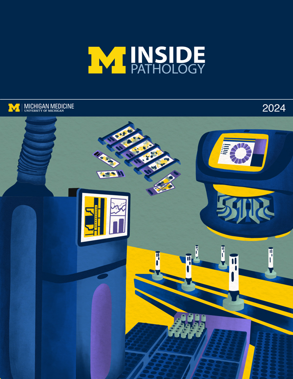
Illustration representing the various machines and processing used within our labs. |
newsletter
INSIDE PATHOLOGYAbout Our NewsletterInside Pathology is an newsletter published by the Chairman's Office to bring news and updates from inside the department's research and to become familiar with those leading it. It is our hope that those who read it will enjoy hearing about those new and familiar, and perhaps help in furthering our research. CONTENTS
|
 ON THE COVER
Rendering of the D. Dan and Betty Khn Health Care Pavilion. Credit: HOK |
newsletter
INSIDE PATHOLOGYAbout Our NewsletterInside Pathology is an newsletter published by the Chairman's Office to bring news and updates from inside the department's research and to become familiar with those leading it. It is our hope that those who read it will enjoy hearing about those new and familiar, and perhaps help in furthering our research. CONTENTS
|
MLabs, established in 1985, functions as a portal to provide pathologists, hospitals. and other reference laboratories access to the faculty, staff and laboratories of the University of Michigan Health System’s Department of Pathology. MLabs is a recognized leader for advanced molecular diagnostic testing, helpful consultants and exceptional customer service.