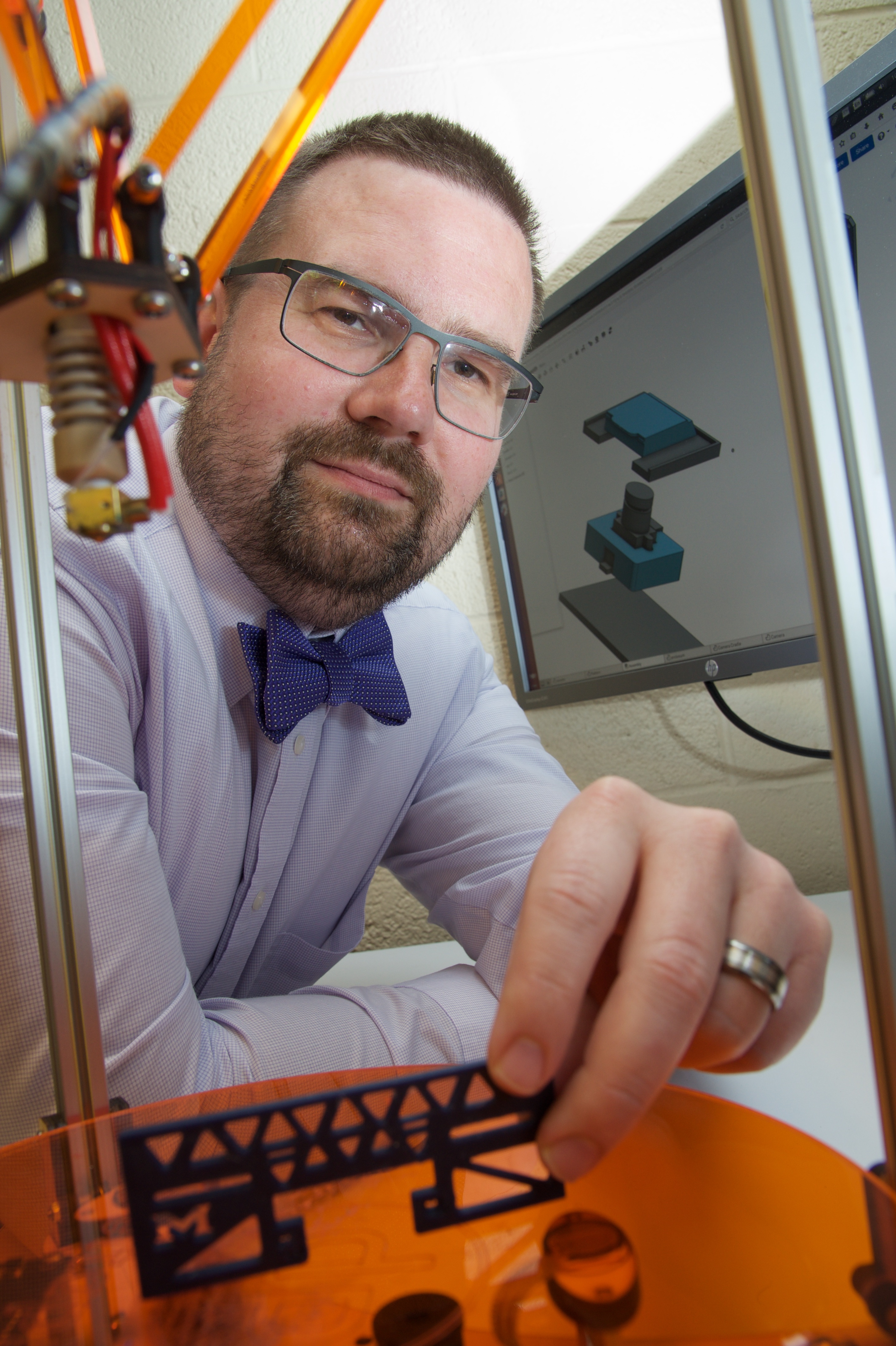
Chris Williams, M.D. examining a custom manufactured part for the prototype histology appliance.
Photography by Elizabeth WalkerDr. Christopher Williams methodically uses a digital caliper to take one final confirmatory measurement of a digital camera module. The value he obtains agrees with his previously rendered engineering diagrams and he then turns his attention to the 3D printer's control console. With a few key clicks, the sleek and futuristic machine stirs into action with its ultra-high precision robotics, depositing one layer of polymer at a time to fabricate an intricate structure. Soon, what was only a computer blueprint will be transformed into a real world physical object - a precision casing for one of the multiple automation instruments now under development within the Pathology Departments's Informatics Division, at the University of Michigan. Chris performs this task with the calm, methodical confidence that one usually only finds in a seasoned design engineer or machinist. This is not unexpected, however, as he is an experienced electrical engineer, with many years of field work under his belt. But what is out of the ordinary is the fact that Dr. Williams is also a Pathologist, and the division's current Informatics fellow.
Nearly a decade ago, despite being a highly successful design engineer, Dr. Williams made a professional decision that few engineers ever arrive upon -- to seek further additional training towards the goal of becoming a physician-engineer. In the US, there are at present only 37 such individuals who identify themselves as combined electrical engineer/pathologists, making Chris part of a very unusual group of designers and inventors. At the conclusion of his residency training at the University of Oklahoma, seeking to further capitalize on his engineering background, Chris made the second unusual career decision to seek further sub-specialty training in pathology informatics, and in so doing, enjoining him to an even smaller guild: only six individuals in the US and perhaps only twice that number worldwide are able to call themselves pathology informaticist-engineers. Indeed, his colleagues joke with him that to simply call him 'one-in-a-million' is a gross underestimate.
Now approaching the close of the first year of his two-year fellowship, Dr. Williams has had the opportunity to extensively canvas the field of pathology informatics and in so doing, he identified a number of areas of critical path workflow for Anatomic Pathology where there remains significant risk for specimen mis-identification. Recognizing that engineered solutions, in the form of compact asset identification appliances, could effectively address these existing workflow deficits, he sought out to design and build the required devices. These devices will house multiple high resolution digital cameras, proximity sensors and make use of sophisticated barcode recognition software, allowing for instant identification of slides and paraffin blocks, as they course their way through the department.
With the completion of the initial design plans, it became evident that any rendered solution would require a significant portfolio of custom-machined or custom-molded parts, or order to realize a device that could reliably withstand the harsh environment as encountered in the modern histology laboratory. A quick accounting of the potential machining and molding expenses quickly placed the project into a prohibitively expensive budget range, even when considering the fabrication of only a small number of prototypes.
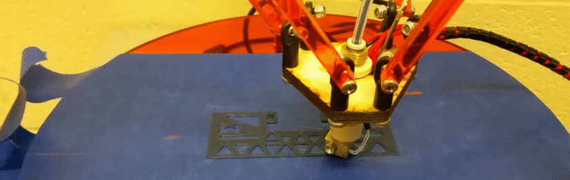
Extrusion assembly of the 3D printer.
Photography by Elizabeth WalkerFacing substantial and potentially intractable fabrication costs, Dr. Williams, being an engineer, did what engineers do very well: he engineered his own solution. Recognizing that 3D printing technology had recently matured to the point where locally manufactured parts can be equal in quality to those fabricated in a dedicated foundry, Chris selected a highly flexible 3D printing solution and then, to save even additional expense, chose to assemble the working unit himself, foregoing the more expensive choice of purchasing a fully assembled model.
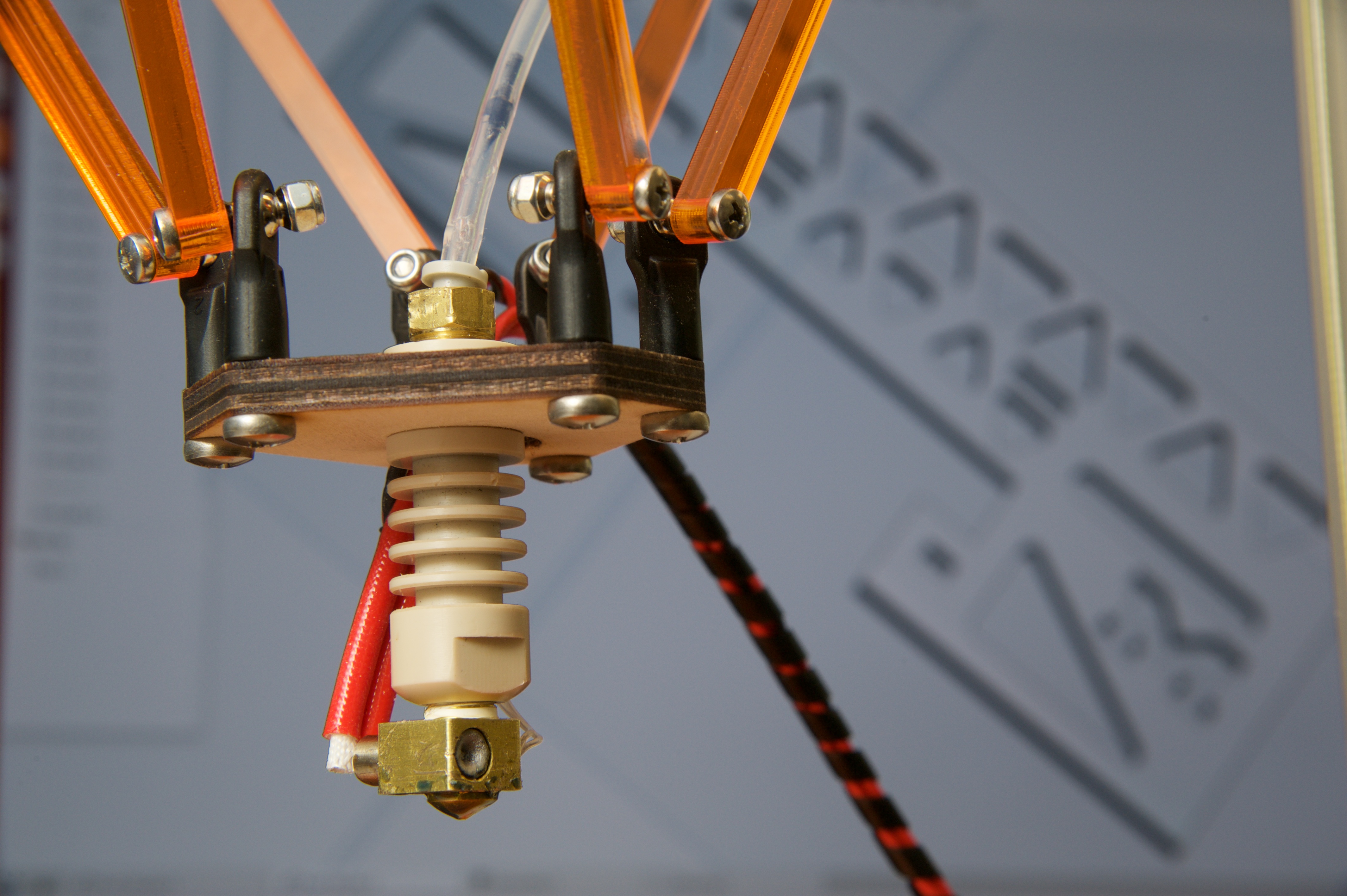
Upon final assembly of the 3D printing unit, Dr. Williams was able to validate that it was operating properly, making it possible for him to fabricate the needed parts in-house. This effort is now well underway. When completed, the finished components will allow for the fabrication of a new class of laboratory automation tool -- tools that allow for simplified tracking of clinical specimens at critical points of processing during their traversal through the histology lab. These specialized appliances will make use of multiple cameras, which will automatically capture images of all sides of a paraffin block, doing so at the same time. And with the newly-gained ability to rapidly fabricate multiple device components locally, it will become possible to construct successive iterations of prototypes, in a fraction of time and at a fraction of the cost of conventional manufacturing techniques.
At the same time, he and Dr. Ulysses Balis, the director of Pathology Informatics, realized that this capability could be of general utility to the department's expansive research community, and have started the process of standing up a core fabrication resource that will be accessible by all members of the Pathology department.
With the remaining time that Dr. Williams has left to complete his two-year fellowship, he plans on fabricating an entire range of such purpose-built appliances, with each unique engineered solution contributing towards the goal of fully automating the anatomic pathology tissue tracking process.
 ON THE COVER
Breast team reviewing a patient's slide. (From left to right) Ghassan Allo, Fellow; Laura Walters, Clinical Lecturer; Celina Kleer, Professor. See Article |
newsletter
INSIDE PATHOLOGYAbout Our NewsletterInside Pathology is an newsletter published by the Chairman's Office to bring news and updates from inside the department's research and to become familiar with those leading it. It is our hope that those who read it will enjoy hearing about those new and familiar, and perhaps help in furthering our research. CONTENTS
|
 ON THE COVER
Autopsy Technician draws blood while working in the Wayne County morgue. See Article |
newsletter
INSIDE PATHOLOGYAbout Our NewsletterInside Pathology is an newsletter published by the Chairman's Office to bring news and updates from inside the department's research and to become familiar with those leading it. It is our hope that those who read it will enjoy hearing about those new and familiar, and perhaps help in furthering our research. CONTENTS
|
 ON THE COVER
Dr. Sriram Venneti, MD, PhD and Postdoctoral Fellow, Chan Chung, PhD investigate pediatric brain cancer. See Article |
newsletter
INSIDE PATHOLOGYAbout Our NewsletterInside Pathology is an newsletter published by the Chairman's Office to bring news and updates from inside the department's research and to become familiar with those leading it. It is our hope that those who read it will enjoy hearing about those new and familiar, and perhaps help in furthering our research. CONTENTS
|
 ON THE COVER
Director of the Neuropathology Fellowship, Dr. Sandra Camelo-Piragua serves on the Patient and Family Advisory Council. |
newsletter
INSIDE PATHOLOGYAbout Our NewsletterInside Pathology is an newsletter published by the Chairman's Office to bring news and updates from inside the department's research and to become familiar with those leading it. It is our hope that those who read it will enjoy hearing about those new and familiar, and perhaps help in furthering our research. CONTENTS
|
 ON THE COVER
Residents Ashley Bradt (left) and William Perry work at a multi-headed scope in our new facility. |
newsletter
INSIDE PATHOLOGYAbout Our NewsletterInside Pathology is an newsletter published by the Chairman's Office to bring news and updates from inside the department's research and to become familiar with those leading it. It is our hope that those who read it will enjoy hearing about those new and familiar, and perhaps help in furthering our research. CONTENTS
|
 ON THE COVER
Dr. Kristine Konopka (right) instructing residents while using a multi-headed microscope. |
newsletter
INSIDE PATHOLOGYAbout Our NewsletterInside Pathology is an newsletter published by the Chairman's Office to bring news and updates from inside the department's research and to become familiar with those leading it. It is our hope that those who read it will enjoy hearing about those new and familiar, and perhaps help in furthering our research. CONTENTS
|
 ON THE COVER
Patient specimens poised for COVID-19 PCR testing. |
newsletter
INSIDE PATHOLOGYAbout Our NewsletterInside Pathology is an newsletter published by the Chairman's Office to bring news and updates from inside the department's research and to become familiar with those leading it. It is our hope that those who read it will enjoy hearing about those new and familiar, and perhaps help in furthering our research. CONTENTS
|
 ON THE COVER
Dr. Pantanowitz demonstrates using machine learning in analyzing slides. |
newsletter
INSIDE PATHOLOGYAbout Our NewsletterInside Pathology is an newsletter published by the Chairman's Office to bring news and updates from inside the department's research and to become familiar with those leading it. It is our hope that those who read it will enjoy hearing about those new and familiar, and perhaps help in furthering our research. CONTENTS
|
 ON THE COVER
(Left to Right) Drs. Angela Wu, Laura Lamps, and Maria Westerhoff. |
newsletter
INSIDE PATHOLOGYAbout Our NewsletterInside Pathology is an newsletter published by the Chairman's Office to bring news and updates from inside the department's research and to become familiar with those leading it. It is our hope that those who read it will enjoy hearing about those new and familiar, and perhaps help in furthering our research. CONTENTS
|
 ON THE COVER
Illustration representing the various machines and processing used within our labs. |
newsletter
INSIDE PATHOLOGYAbout Our NewsletterInside Pathology is an newsletter published by the Chairman's Office to bring news and updates from inside the department's research and to become familiar with those leading it. It is our hope that those who read it will enjoy hearing about those new and familiar, and perhaps help in furthering our research. CONTENTS
|
 ON THE COVER
Rendering of the D. Dan and Betty Khn Health Care Pavilion. Credit: HOK |
newsletter
INSIDE PATHOLOGYAbout Our NewsletterInside Pathology is an newsletter published by the Chairman's Office to bring news and updates from inside the department's research and to become familiar with those leading it. It is our hope that those who read it will enjoy hearing about those new and familiar, and perhaps help in furthering our research. CONTENTS
|
MLabs, established in 1985, functions as a portal to provide pathologists, hospitals. and other reference laboratories access to the faculty, staff and laboratories of the University of Michigan Health System’s Department of Pathology. MLabs is a recognized leader for advanced molecular diagnostic testing, helpful consultants and exceptional customer service.