Qualitative Image Analysis study shows excellent results
By Lynn McCain | June 9 2022A  landmark study into quantitative image analysis in ER, PgR, and HER2 in invasive breast carcinoma was recently published in the American Journal of Clinical Pathology. Dr. Mustafa Yousif, Assistant Professor of Breast Pathology and Informatics, and colleagues conducted a retrospective study of 1,367 invasive breast carcinomas of all histopathology subtypes, for which ER, PgR, and HER2 were analyzed by manual scoring. These were compared to the results obtained using quantitative image analysis (QIA). QIA uses a form of artificial intelligence (AI) called deep learning to identify specific regions of interest and to interpret that based on programmed algorithms.
The team found concordance between QIA and manual scores for ER, PgR, and HER2 was 93%, 96%, and 90% respectively. Discordant cases had low positive scores (1%-10%) for ER (n=33), were due to nonrepresentative region selection (e.g., ductal carcinoma in situ) or tumor heterogeneity for PgR (n=43), and were of one-step difference (negative to equivocal, equivocal to positive, or vice versa) for HER2 (n=90). Among HER2 cases where FISH results were available, only four (1%) showed discordant QIA and FISH results. Overall, use of QIA significantly improves the standardization of scoring of biomarker expression evaluations and demonstrated excellent concordance with pathologists’ scores. While pathologist oversight of representative region selection is still advised, this is another important step in advancing the practice of pathology to ensure the best possible patient outcomes.
Today, at Michigan Medicine, we accordingly analyze all new breast carcinoma cases using QIA.
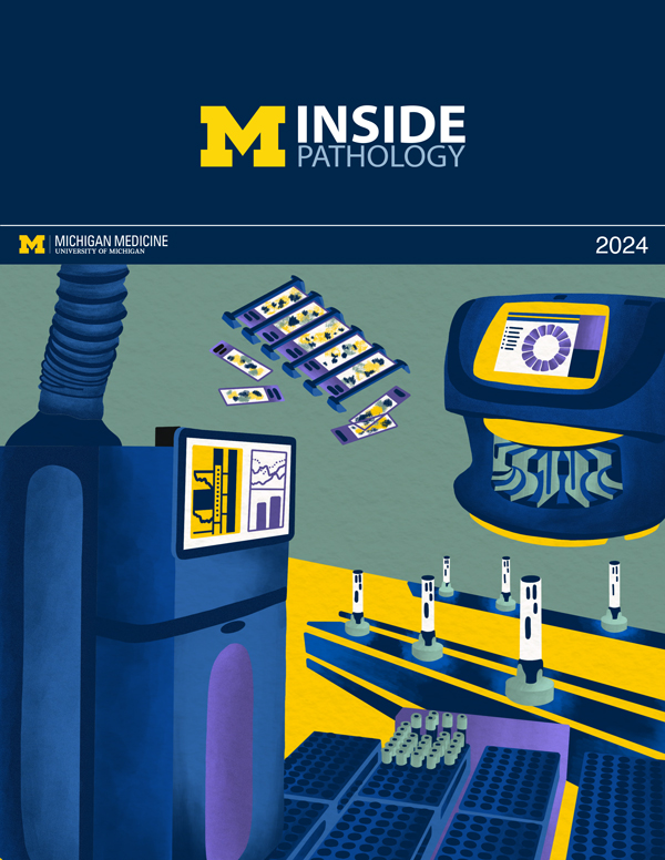
 ON THE COVER
 ON THE COVER
 ON THE COVER
 ON THE COVER
 ON THE COVER
 ON THE COVER
 ON THE COVER
 ON THE COVER
 ON THE COVER
 ON THE COVER
 ON THE COVER