2024 Case StudiesExplore the cases that will be discussed at this year's conference. Here you'll find the Case History of each with links to their biopsies. Quick Jump
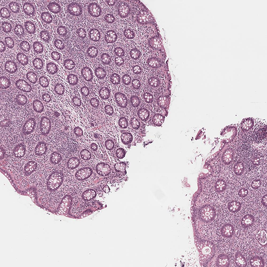
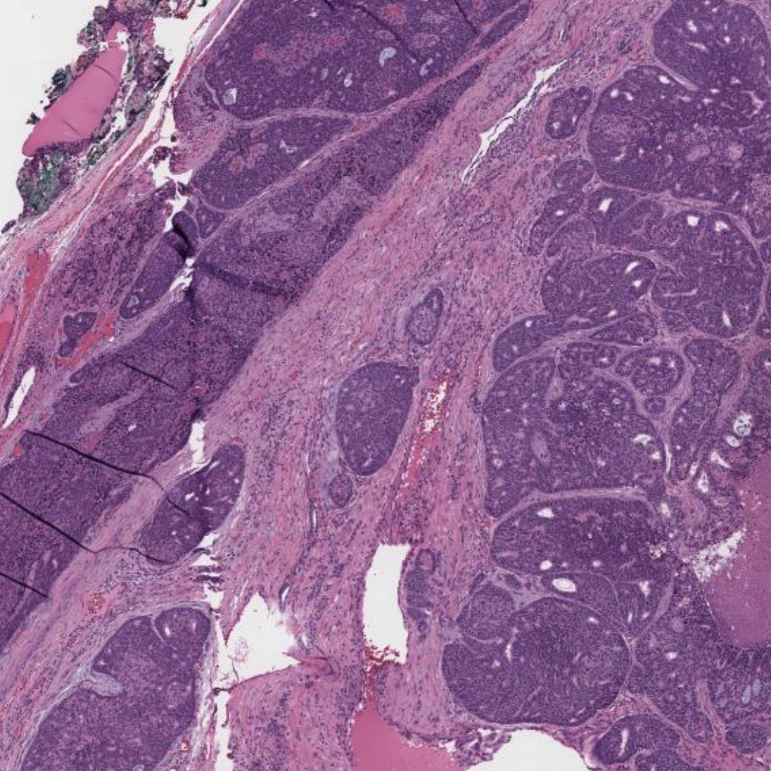
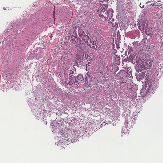
Case History / 02Ellen Chapel, MD / Breast Pathology 54-year-old female with a left breast mass, described by initial imaging as a 2.7 x 1.6 x 2.1 cm hypoechoic lesion at the 2:00 position. Biopsy demonstrated poorly differentiated invasive ductal carcinoma (ER-low, PR-negative, HER2-negative). She was treated with neoadjuvant chemotherapy and now presents for surgical management. Case History / 05Margaret Fang, MD / GI Pathology A 56-year-old male has a history of primary biliary cholangitis and chronic diarrhea of 12 years.
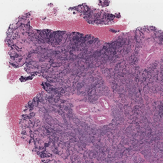
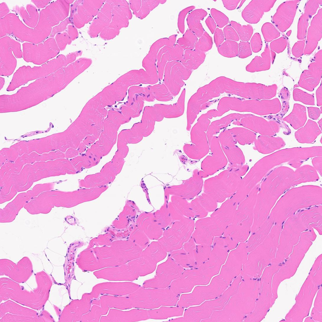
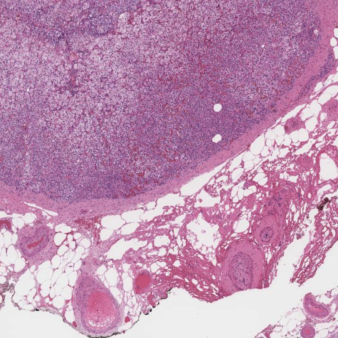
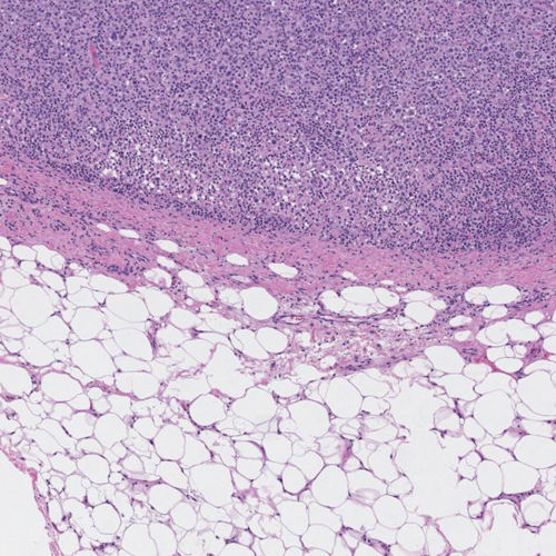
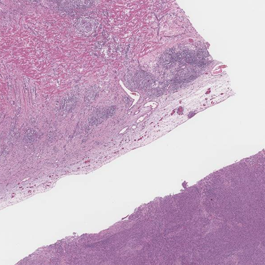
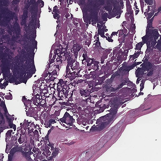
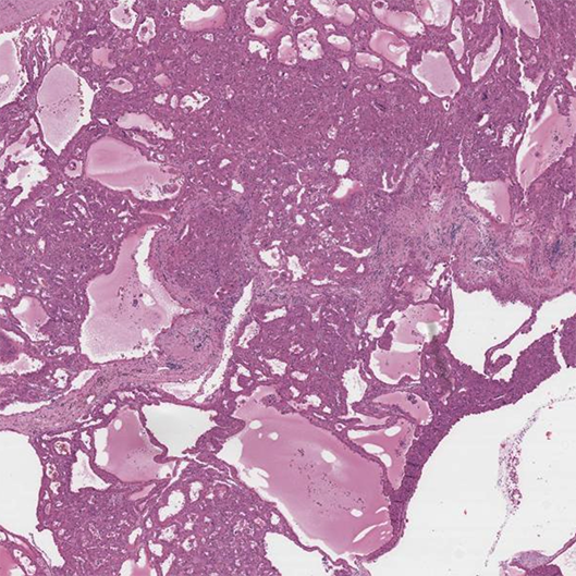
Case History / 06Kyle Perry, MD / Bone & Soft Tissue Pathology 45-year-old male with a large mass in the retroperitoneum. The tumor was excised. Case History / 07Mark Girton, MD / Hematopathology A 70-year-old male presented to a dental clinic for a one-year history of non-healing necrotic gingival ulcer, 1.2 cm in greatest dimension, after extensive dental work for poor dentition. The clinical differential diagnosis included necrotic ulcerative periodontitis versus oral squamous cell carcinoma. Of note, the patient has a 12-year history of chronic lymphocytic leukemia(CLL) with observation only. Case History / 08Sean Ferris, MD, PhD / Neuropathology A now 58-year-old man began having progressive proximal muscle weakness in June, 2023. Subsequently also developed dysphagia to dry foods (cutting small bites), shortness of breath with exertion, and weight loss due to low appetite. No skin rashes noted. Serum creatine kinase level was 7,200 IU/L (upper limit of normal is 240 IU/L). Electromyography (EMG) study demonstrated evidence of an active irritable myopathy affecting proximal more than distal muscles. Patient underwent left deltoid muscle biopsy in January, 2024. Case History / 09Aaron Udager, MD, PhD / Endocrine Pathology A 51-year-old woman with a history of resistant hypertension and an incidentally identified 1.3 cm left adrenal gland nodule. Pre-operative laboratory testing revealed hypokalemia and hyporeninemic hyperaldosteronism, and adrenal venous sampling showed lateralization of excess aldosterone to the left adrenal vein. A left adrenalectomy was subsequently performed. Case History / 11Xiaobing Jin, MD, PhD / Cytopathology A 62-year-old female presented with a 1.2 cm mass-like lesion in left submandibular gland. ACT scan revealed interval development of a focal hypoenhancement of the mass. The differential diagnoses for this CT findings include neoplastic and inflammatory changes. No neck lymphadenopathy was noted. Patient is a never smoker. Case History / 12Jaclyn Plotzke, MD / Dermatopathology A 51-year-old male with metastatic esophageal carcinoma on pembrolizumab. The patient had been tolerating multiple rounds of pembrolizumab with mild cutaneous eruptions in the past. The clinical team added carboplatin to his regimen, after which he developed a rash progressing from maculopapular to vesiculobullous (involving >70% total body surface area with mucous membrane involvement) over the course of weeks. Case History / 13Tao Huang, BM, PhD / Thoracic Pathology 73-year-old man who presented with chest pain was incidentally found to have a 2.5 cm lung mass on CT angiogram. The lung nodule is FDG avid with no evidence of regional nodal or distant metastasis on PET. He is physically fit and a non-smoker. A diagnostic transbronchial biopsy was performed. Case History / 15Kristine Konopka, MD / Thoracic Pathology Along with patient demographics (not provided here), the requisition only lists the specimen as “Right lung cryobiopsy” and preoperative diagnosis as “ILD.”
Break Out Cases
Break Out Case History / 01Kamran Mirza, MBBS, PhD / Hematopathology
Break Out Case History / 02Maria Westerhoff, MD / GI Pathology
Break Out Case History / 03Jeffrey Myers, MD & Heather Chen-Yost, MD / Thoracic Pathology Break Out Case History / 04Eman Abdulfatah, MBBch, MSc & Angela Wu, MD / GU Pathology
|


